
Page 961 - Cote clinical veterinary advisor dogs and cats 4th
P. 961
Hip Dysplasia 471
VetBooks.ir Diseases and Disorders
A B C D
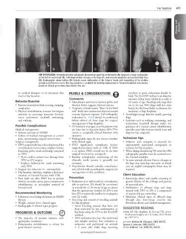
HIP DYSPLASIA Ventrodorsal pelvic radiographs demonstrate good hip conformation (A), dysplastic change (subluxation
of the left femoral head) (B), mild degenerative changes in the hips (C), and severely dysplastic and osteoarthritic hips
(D). Radiographic abnormalities (D) include severe subluxation of the femoral heads and remodeling of the shallow
acetabula and femoral necks. This dog would be a candidate for total hip replacement or femoral head/neck ostectomies,
based on clinical parameters described in the text.
to medical therapies or in situations that PEARLS & CONSIDERATIONS excellent or good evaluations should be
involve hip luxation. bred. The PennHIP method is an objective
Comments measure of hip laxity validated as early as
Behavior/Exercise • Musculature asymmetry between pelvic and 16 weeks of age. Breeding only dogs that
• Exercise moderation; limit running, jumping, thoracic limbs suggests bilateral disease. are in the top 50% (dogs with less joint
rough play • Dysplastic animals acutely “down in the hind laxity) for the breed helps to decrease the
• Physical rehabilitation; increase low-impact end” likely have concurrent bilateral cranial incidence of hip dysplasia.
activities to encourage muscular develop- cruciate ligament rupture. Full orthopedic • Avoid high-energy diets for rapidly growing
ment: underwater treadmill, swimming, evaluation (p. 1143) should be performed dogs.
and walking before referral of these dogs for surgical • Activities such as walking, swimming, and
management of hip dysplasia. underwater treadmill therapy under the
Possible Complications • Of medically managed juvenile patients that guidance of a trained canine rehabilitation
Medical management: are lame due to hip joint laxity, 60%-75% specialist may help increase muscle mass and
• Adverse reaction to NSAID return to acceptable clinical function with improve hip congruity.
• Failure of medical management to control maturity.
pain, necessitating surgical intervention • Radiographic signs do not always correlate Technician Tips
Surgical management: with clinical signs. • Sedation with analgesia is required for
• DPO purportedly has reduced potential for • FHO significantly complicates further appropriately positioned radiographs to
complications (nerve injury, implant failure/ surgical procedures such as THR. If THR evaluate for hip dysplasia.
loosening, pelvic canal narrowing) compared is an option, FHO should not be the first • When taking extended-leg VD views for OFA
to TPO. surgical intervention attempted. radiographs, patellas must be centered over
○ Nerve and/or urinary tract damage from • Routine radiographic monitoring of the the femoral condyles.
TPO or JPS surgery clinically stable patient is generally not • In some animals chronic fibrotic changes in
○ Implant failure/pelvic canal narrowing necessary. the hips may limit hip extension, in which
with TPO • Practitioners should consider consultation case an angled radiographic beam may be
• Surgical site infection +/− osteomyelitis and referral with an orthopedic surgeon for required.
• Hip luxation, infection, implant subsidence/ management of this condition.
rotation, or femoral fracture with THR Client Education
• Poor limb use after FHO due to patient Prevention • Knowledge about and careful screening of
obesity, muscular atony, poor postoperative • Evaluation of at-risk breeds by a veterinarian genetic history of the siblings and parents
rehabilitation, or incomplete removal of able to perform JPS should be considered of potential pets
femoral neck in animals by 14-16 weeks of age to ensure • Sterilization of affected dogs and dogs
that the appropriate window for JPS is not treated with TPO or JPS is a component
Recommended Monitoring lost; JPS is reportedly ineffective in puppies of responsible pet ownership.
• Laboratory monitoring of patients on NSAID over 22 weeks. • Minimize clinical signs in affected dogs
therapy • Screening and control of breeding animals through diet, low-impact exercise (see
• Weight, exercise level, clinical signs for hip dysplasia Prevention above), and medical management.
• Radiographs if clinical signs progress ○ Avoid breeding animals that have not
been evaluated for hip conformation or SUGGESTED READING
PROGNOSIS & OUTCOME hip laxity using the OFA or the PennHIP Schulz KS: Hip dysplasia. In Fossum TW, editor:
method. Small animal surgery, ed 4, St. Louis, 2013, Mosby,
• The majority of patients improve with ○ OFA evaluation has been the traditional pp 1305-1316.
appropriate treatment. and simpler method, but evaluation is AUTHORS: Mathieu M. Glassman, VMD, DACVS;
• Postoperative rehabilitation is critical for subjective and performed on animals Spencer A. Johnston, VMD, DACVS
good clinical recovery. > 2 years old. Only dogs receiving EDITOR: Kathleen Linn, DVM, MS, DACVS
www.ExpertConsult.com