
Page 963 - Cote clinical veterinary advisor dogs and cats 4th
P. 963
472 Hip Luxation
Hip Luxation Client Education
Sheet
VetBooks.ir
BASIC INFORMATION
• With craniodorsal luxation, the limb appears
shortened, with stifle rotated laterally and • Limb and pelvic exams plus orthogonal
view radiography to confirm the diagnosis
Definition calcaneus medially. and determine concomitant or inciting
• Dislocation of the femoral head relative to the • Palpation of hip reveals swelling, pain, disorders
acetabulum, most frequently in a craniodorsal crepitus, and abnormal position of greater
direction trochanter. TREATMENT
• Luxation: complete displacement of the
femoral head relative to the acetabulum Etiology and Pathophysiology Treatment Overview
• Subluxation: partial or incomplete dis- Coxofemoral luxations comprise 90% of all • With acute injury of a normal hip joint,
location of the femoral head relative to luxations in small animals. the goal is closed or open anatomic joint
the acetabulum (i.e., joint incongruity • With craniodorsal luxation, femoral head is reduction/stabilization.
exists) driven dorsally over the acetabular rim. • With an abnormal or chronically luxated
○ The femoral head ligament and joint normal joint, the goal is to return pain-free
Synonyms capsule are torn. function with femoral head and neck exci-
Coxofemoral luxation, dislocated hip ○ Avulsion fracture associated with the sion (FHNE), also known as femoral head
ligament can occur. ostectomy (FHO), total hip replacement
Epidemiology ○ Gluteal muscle contractions exacerbate (THR), or pelvic osteotomy.
SPECIES, AGE, SEX craniodorsal displacement of proximal femur.
Dogs and cats of either sex and any age • With ventral luxation, transverse acetabular Acute General Treatment
ligament is ruptured. • Closed reduction (p. 1158) and Ehmer sling
RISK FACTORS application (p. 1161) if
Dogs with hip dysplasia are at risk for luxation DIAGNOSIS ○ Duration of injury is < 48 hours and
with even minor trauma. ○ Hip is otherwise structurally normal and
Diagnostic Overview ○ Patient is sufficiently stable to undergo
ASSOCIATED DISORDERS Orthogonal view radiography establishes the general anesthesia
Pelvic and proximal femoral fractures diagnosis and shows any concomitant fractures • For craniodorsal luxation with patient in
in the region. lateral recumbency (unaffected side down),
Clinical Presentation the upper (affected) limb is externally rotated
DISEASE FORMS/SUBTYPES Differential Diagnosis (toes out), abducted (lifted, as a male dog
Craniodorsal luxation is most common; Subluxation due to hip dysplasia, femoral lifts the leg to urinate), and then internally
caudoventral is uncommon. capital physeal fracture, femoral neck fracture, rotated (toes in) while manual pressure on
acetabular fracture trochanter guides head into acetabulum.
HISTORY, CHIEF COMPLAINT • After reduction, extensive range-of-motion
• Trauma (falls, vehicular) causing acute Initial Database maneuvers are performed to force soft tissue
non–weight-bearing lameness • Physical exam (see Physical Exam Findings out of acetabulum and test joint stability.
• Intermittent lameness in chronic cases with above) • Open (surgical) reduction is performed
unrecognized trauma • CBC, serum biochemistry panel, and uri- for failed or unstable closed reductions in
nalysis to assess anesthetic risk (American patients with normal coxofemoral joints:
PHYSICAL EXAM FINDINGS Society of Anesthesiologists classification ○ Extraarticular techniques: capsulorrhaphy,
• Variable lameness system) (p. 1196) suture between origin of greater trochanter
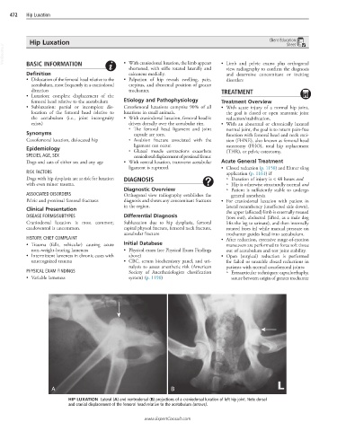
A B
HIP LUXATION Lateral (A) and ventrodorsal (B) projections of a craniodorsal luxation of left hip joint. Note dorsal
and cranial displacement of the femoral head relative to the acetabulum (arrows).
www.ExpertConsult.com