
Page 819 - Withrow and MacEwen's Small Animal Clinical Oncology, 6th Edition
P. 819
CHAPTER 34 Miscellaneous Tumors 797
longer than those not having RT with an MST of 182 days. 571
Dogs treated with a set combined protocol of a palliative RT and
CCNU had an MST of 208 days. Further study into the optimal
VetBooks.ir RT protocol for HS is necessary.
Hemophagocytic Histiocytic Sarcoma
Hemophagocytic HS is a variant of HS that originates from the
tissue macrophage, not the DC. 483 This more aggressive form of
HS invariably involves the spleen, but dogs may also have liver,
bone marrow, LN, and/or lung involvement. Splenic involve-
ment is usually diffuse, resulting in gross enlargement with diffuse
nodular infiltrates. In one study, common hematologic findings in
dogs with HHS included a regenerative anemia (94%), thrombo-
cytopenia (88%), hypoalbuminemia (94%), and hypocholesterol-
emia (69%). 483 A presumptive diagnosis of HHS may be obtained
through splenic cytology, which shows infiltration with atypical
to highly pleomorphic macrophages displaying phagocytosis
of red blood cells, splenic origin red cell precursors, and white
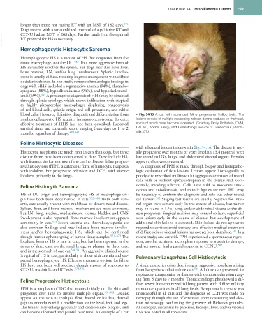
blood cells. However, definitive diagnosis and differentiation from • Fig. 34.16 A cat with advanced feline progressive histiocytosis. The
nonhemophagocytic HS requires immunophenotyping. To date, lesions consist of multiple coalescing hairless dermal nodules on the head,
effective treatment of HHS has not been described. Reported some of which have become ulcerated. (Courtesy Emily Rothstein, DVM,
survival times are extremely short, ranging from days to 1 to 2 DACVD, Animal Allergy and Dermatology Service of Connecticut, Plants-
months, regardless of therapy. 483,542 ville, CT.)
Feline Histiocytic Diseases
with advanced lesions in shown in Fig. 34.16. The disease is usu-
Histiocytic neoplasms are much rarer in cats than dogs, but three ally progressive over months or years (median 13.4 months) with
distinct forms have been documented to date. These include HS, late spread to LNs, lungs, and abdominal visceral organs. Females
with features similar to those of the canine disease; feline progres- appear to be overrepresented.
sive histiocytosis (FPH), a cutaneous form of histiocytic neoplasia A diagnosis of FPH is made through biopsy and histopatho-
with indolent, but progressive behavior; and LCH, with disease logic evaluation of skin lesions. Lesions appear histologically as
localized primarily to the lungs. poorly circumscribed multinodular aggregates or masses of round
cells with or without epitheliotropism in the dermis and, occa-
Feline Histiocytic Sarcoma sionally, invading subcutis. Cells have mild to moderate aniso-
cytosis and anisokaryosis, and mitotic figures are rare. IHC may
HS of DC origin and hemophagocytic HS of macrophage ori- be necessary to confirm the diagnosis and rule out other round
gin have both been documented in cats. 572–580 With both vari- cell tumors. 582 Staging test results are usually negative for inter-
ants, cats usually present with multifocal or disseminated disease. nal organ involvement early in the course of disease, but tumor
Spleen, liver, and bone marrow involvement are most common, may be found in LNs, lung, and/or abdominal viscera as the dis-
but LN, lung, trachea, mediastinum, kidney, bladder, and CNS ease progresses. Surgical excision may control solitary, superficial
involvement is also reported. Bone marrow involvement appears skin lesions early in the course of disease, but development of
commonly in cats. 573 Severe anemia and thrombocytopenia are additional skin lesions is expected. Skin lesions do not appear to
also common findings and may indicate bone marrow involve- respond to corticosteroid therapy, and effective medical treatment
ment and/or hemophagocytic HS, which can be confirmed of diffuse skin or visceral lesions has not yet been described. 581 In a
though immunophenotyping of tumor tissue samples. 571–573 The recent study, one cat with FPH experienced a spontaneous regres-
localized form of HS is rare in cats, but has been reported in the sion, another achieved a complete response to masitinib therapy,
tarsus of three cats, on the nasal bridge or planum in three cats, and yet another had a partial response to CCNU. 582
and in the stomach of one cat. 580,581 An aggressive clinical course
is typical of HS in cats, particularly in those with anemia and sus- Pulmonary Langerhans Cell Histiocytosis
pected hemophagocytic HS. Effective treatment options for feline
HS have not been well studied, though reports of responses to A single case series exists describing an aggressive neoplasm arising
CCNU, masinitib, and RT exist. 574,581 from Langerhans cells in three cats. 583 All three cats presented for
respiratory compromise or distress with symptom duration rang-
Feline Progressive Histiocytosis ing from 5 days to 7 months. Thoracic radiographs showed a dif-
fuse, severe bronchointerstitial lung pattern with diffuse military
FPH is a neoplasm of DC that occurs initially on the skin and to nodular opacities in all lung fields. Symptomatic therapy was
progresses over time to involve multiple organs. 581,582 Lesions unsuccessful in all cats and the diagnosis of LCH was made on
appear on the skin as multiple firm, haired or hairless, dermal necropsy through the use of extensive immunostaining and elec-
papules or nodules with a predilection for the head, feet, and legs. tron microscopy confirming the presence of Birbeck’s granules.
The lesions may enlarge gradually and coalesce into plaques, and At necropsy, metastasis to pancreas, kidneys, liver, and/or visceral
can become ulcerated and painful over time. An example of a cat LNs was noted in all three cats. 