
Page 814 - Withrow and MacEwen's Small Animal Clinical Oncology, 6th Edition
P. 814
792 PART IV Specific Malignancies in the Small Animal Patient
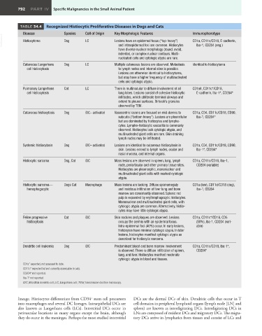
TABLE 34.4 Recognized Histiocytic Proliferative Diseases in Dogs and Cats
Disease Species Cell of Origin Key Morphologic Features Immunophenotype
VetBooks.ir Histiocytoma Dog LC Lesions have an epidermal focus (“top-heavy”) CD1a, CD11c/CD18, E-cadherin,
and intraepidermal foci are common. Histiocytes
Iba-1, CD204 (neg.)
have diverse nuclear morphology (round, ovoid,
indented, or complex nuclear contours. Multi-
nucleated cells and cytologic atypia are rare.
Cutaneous Langerhans Dog LC Multiple cutaneous lesions are observed. Metastasis Identical to histiocytoma
cell histiocytosis to lymph nodes and internal sites is possible.
Lesions are otherwise identical to histiocytoma,
but may have a higher frequency of multinucleated
cells and cytologic atypia.
Pulmonary Langerhans Cat LC There is multinodular to diffuse involvement of all CD1a#, CD11c*/CD18,
cell histiocytosis lung lobes. Lesions consist of cohesive histiocytic E-cadherin, Iba-1*, CD204*
infiltrates, which obliterate terminal airways and
extend to pleural surfaces. Birbeck’s granules
observed by TEM.
Cutaneous histiocytosis Dog iDC– activated Vasocentric lesions are focused on mid-dermis to CD1a, CD4, CD11c/CD18, CD90,
subcutis (“bottom heavy”). Lesions are pleocellular Iba-1, CD204*
but are dominated by histiocytes and lympho-
cytes. Lympho-histiocytic vasculitis is commonly
observed. Histiocytes lack cytologic atypia, and
multinucleated giant cells are rare. Skin draining
lymph nodes may be infiltrated.
Systemic histiocytosis Dog iDC– activated Lesions are identical to cutaneous histiocytosis in CD1a, CD4, CD11c/CD18, CD90,
skin. Lesions extend to lymph nodes, ocular and Iba-1*, CD204*
nasal mucosa, and internal organs.
Histiocytic sarcoma Dog, Cat iDC Mass lesions are observed in spleen, lung, lymph CD1a, CD11c/CD18, Iba-1,
node, periarticular and other primary tissue sites. CD204 (variable)
Histiocytes are pleomorphic, mononuclear and
multinucleated giant cells with marked cytologic
atypia.
Histiocytic sarcoma— Dogs Cat Macrophage Mass lesions are lacking. Diffuse splenomegaly CD1a (low), CD11d/CD18 (dog),
hemophagocytic and insidious infiltration of liver lung and bone iba-1, CD204
marrow are consistently observed. Splenic red
pulp is expanded by erythrophagocytic histiocytes.
Mononuclear and multinucleated giant cells, with
cytologic atypia are common. Alternatively, histio-
cytes may have little cytologic atypia.
Feline progressive Cat iDC Skin nodules and plaques are observed. Lesions CD1a, CD11c*/CD18, CD5
histiocytosis occupy the dermis with an epidermal focus. (50%), Iba-1, CD204 (vari-
Intra-epidermal foci (40%) occur. In early lesions, able)
histiocytes have minimal cytologic atypia. In later
lesions, histiocytes manifest cytologic atypia as
described for histiocytic sarcoma.
Dendritic cell leukemia Dog iDC Predominant blood and bone marrow involvement CD1a, CD11c/CD18, Iba-1*,
is observed. There is diffuse infiltration of spleen, CD204*
lung, and liver. Histiocytes manifest moderate
cytologic atypia in blood and tissues.
CD1a* expected, not assessed to date.
CD11c* expected but not currently assessable in cats.
CD204* not reported.
Iba-1* not reported.
iDC, Interstitial dendritic cell; LC, Langerhans cell; TEM, transmission electron microscopy.
lineage. Histiocytes differentiate from CD34 stem cell precursors DCs are the dermal DCs of skin. Dendritic cells that occur in T
+
into macrophages and several DC lineages. Intraepithelial DCs are cell domains in peripheral lymphoid organs (lymph node [LN] and
also known as Langerhans cells (LCs). Interstitial DCs occur in spleen) are known as interdigitating DCs. Interdigitating DCs in
perivascular locations in many organs except the brain, although LNs are composed of resident DCs and migratory DCs. The migra-
they do occur in the meninges. Perhaps the most studied interstitial tory DCs arrive in lymphatics from tissues and consist of LCs and