Page 810 - Withrow and MacEwen's Small Animal Clinical Oncology, 6th Edition
P. 810
788   PART IV    Specific Malignancies in the Small Animal Patient


         be benign or malignant, with the latter predominating, and most   Diagnostic Techniques and Workup
         occur in the right atrium and auricle in the dog. 323,329  Cardiac   Differential diagnosis of a cardiac tumor is often made based on
         HSA is frequently associated with hemorrhagic pericardial effu-
  VetBooks.ir  sion and cardiac tamponade and tends to have high rates of   clinical history, physical examination, and radiographic find-
                          ABTs are the second most common  pri-
                                                               ings. Diagnosis requires imaging, which is routinely achieved by
         metastasis.
                  330,332,347
         mary neoplasm of the heart in dogs, and lymphoma and ecto-  echocardiography  and  sometimes  through  additional  advanced
         pic thyroid carcinoma are observed with some frequency as   modalities such as CT or MRI. Incidental diagnosis of subclinical
         well. 323–326,329,348–359  Reported but rare malignant tumors include   cardiac tumors may be encountered at necropsy. In the major-
         mesothelioma, myxosarcoma, chondrosarcoma, fibrosarcoma,   ity of cases, antemortem cytologic or histologic confirmation of
         osteosarcoma,  rhabdomyosarcoma,  undifferentiated  sarcoma,   neoplasia is not obtained; however, in cases where technically fea-
         leiomyosarcoma, thyroid carcinosarcoma, peripheral nerve sheath   sible and with acceptable clinical risk, FNA or biopsy may provide
         tumor, granular cell tumor, malignant mesenchymoma, and ana-  importance guidance on therapeutic options. 422
         plastic carcinoma. 360–385  Histologically benign cardiac tumors   Much like clinical signs, physical exam findings vary widely
         have been reported and may cause lifethreatening clinical signs   depending on location and hemodynamic consequences of the
         because of their location despite their biologically benign behav-  cardiac tumor. Patients with incidentally identified cardiac tumors
         ior. Reported benign cardiac tumors include myxoma, lipoma,   may have apparently normal physical examinations. Auscultatory
         thyroid  adenoma,  hamartoma,  Schwannoma,  leiomyoma,  and   abnormalities are common secondary to pericardial effusion and
         fibroma. 359,386–399  In contrast to those in humans, metastatic   include muffled heart sounds, pericardial friction rubs, or tumor
         cardiac tumors are diagnosed with less frequency than primary   plops (intermittent diastolic sounds secondary to tumor motion).
         tumors in dogs, likely due to the high incidence of cardiac HSA   In  addition,  arrhythmias  may  be  auscultated,  particularly  in
         in this species and absence of cardiac specific clinical signs for   patients with myocardial involvement. Pulmonary auscultation
         many secondary tumors. 323,326,328  Tumors reported to metastasize   may  reveal abnormalities  consistent  with  left-sided  congestive
         to the heart in dogs include HSA, lymphoma, mammary gland   heart failure, such as increased bronchovesicular sounds and/or
         carcinoma, melanoma, pheochromocytoma, histiocytic sarcoma,   soft crackles. Pulse quality derangements are common in patients
         gastric adenocarcinoma, liposarcoma, malignant mesenchymoma,   in low-output states, such as cardiac tamponade. Jugular venous
         rhabdomyosarcoma,  extraskeletal  osteosarcoma,  fibrosarcoma,   distention and pulsation may also be observed secondary to ele-
         and pulmonary carcinoma. 326,328,400–412              vated right heart pressure with pericardial effusion or obstructive
            Cardiac tumors in cats tend to be malignant although benign   lesions.
         intrapericardial cysts have been reported. 413,414  In addition to lym-  Many  components  of  the  diagnostic  evaluation  are  related
         phoma, ABT, and HSA, single case reports of primary cardiac ganglio-  to the common concomitant condition of pericardial effusion.
         neuroma, rhabdomyosarcoma, and myxoma exist. 415–417  Metastatic   An electrocardiogram may be normal in patients with cardiac
         lesions have been reported to arise from squamous cell carcinoma,   tumors or may show a wide variety of cardiac arrhythmias,
         mammary gland carcinoma, and pulmonary carcinoma. 326,418    which are frequently related to the site of the cardiac tumor and
                                                               infiltration of the myocardium. 423  Conduction disturbances,
         History and Clinical Signs                            such as atrioventricular blocks or bundle branch blocks, may
                                                               be observed with myocardial infiltration and may be as severe
         Tumors of the heart generally cause clinical signs secondary   as complete atrioventricular block as previously reported with
         to  alterations  of  cardiac  function  and  may  result  from  a  mass   cardiac lymphoma. 349  Supraventricular or ventricular arrhyth-
         obstructing  blood flow  to and from  the heart, external  cardiac   mias may be observed in cases of cardiac tumors with or without
         compression that impedes filling such as pericardial effusion, and/  pericardial effusion. ST segment changes may be observed sec-
         or arrhythmias or decreased contractility resulting from myocar-  ondary to myocardial ischemia with or without pericardial effu-
         dial infiltration or ischemia of the myocardium. Clinical signs are   sion. 424  Sinus tachycardia is common with cardiac tamponade
         influenced more by the tumor location, tumor size, and presence   or in cases with heart failure acquired secondary to obstructive
         of pericardial effusion than the specific histology of the tumor.   cardiac tumors.
         Sudden death may occur secondary to cardiac arrhythmias or
         tumor rupture and subsequent blood loss, with or without car-
         diac tamponade. Tumors, particularly cardiac HSA, arising in the
         right side of the heart often cause signs of right-sided congestive
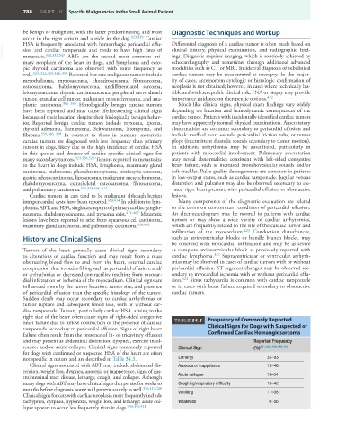
         heart failure due to inflow obstruction or the presence of cardiac   TABLE 34.3     Frequency of Commonly Reported
         tamponade secondary to pericardial effusion. Signs of right heart   Clinical Signs for Dogs with Suspected or
         failure often result from the presence of bi- or tricavitary effusion   Confirmed Cardiac Hemangiosarcoma
         and may present as abdominal distention, dyspnea, exercise intol-                   Reported Frequency
         erance, and/or acute collapse. Clinical signs commonly reported   Clinical Sign     (%) 331,332,458,459,462
         for dogs with confirmed or suspected HSA of the heart are often
         nonspecific in nature and are described in Table 34.3.   Lethargy                        35–93
            Clinical signs associated with ABT may include abdominal dis-  Anorexia or inappetence  19–46
         tension, weight loss, dyspnea, anorexia or inappetence, signs of gas-
         trointestinal tract disease, lethargy, cough, and collapse. Although   Acute collapse    13–54
         many dogs with ABT may have clinical signs that persist for weeks to   Coughing/respiratory difficulty  13–42
         months before diagnosis, some will present acutely as well. 336,419,420
         Clinical signs for cats with cardiac neoplasia most frequently include   Vomiting        11–38
         tachypnea, dyspnea, hyporexia, weight loss, and lethargy; acute col-  Weakness            0–56
lapse appears to occur less frequently than in dogs. 339,345,421 

