
Page 808 - Withrow and MacEwen's Small Animal Clinical Oncology, 6th Edition
P. 808
786 PART IV Specific Malignancies in the Small Animal Patient
not found. Sclerosing mesothelioma must be distinguished from
chronic inflammatory diseases of the body cavity, such as chronic
peritonitis, and histologic examination of biopsy material is essen-
VetBooks.ir tial to establish the diagnosis. Embolized, nonneoplastic mesothe-
lial cells within lymph nodes is a rare finding in humans with
cavity effusions and has been reported in dogs affected with idio-
pathic hemorrhagic pericardial effusion; care must be taken to not
overinterpret these cells as indicative of metastasis. 302
The most useful criteria in establishing a diagnosis of mesothe-
lioma are that the tumor is primarily a neoplasm of the coelomic
cavity lining and that the method of tumor spread is by transcoe-
lomic implantation. Therefore mesothelioma should be consid-
ered when the bulk of the neoplastic tissue exists on the coelomic
surface. Histologically, mesotheliomas need to be differentiated
from carcinomas, adenocarcinomas, or sarcomas, depending on
the morphologic type of the mesothelioma. Unfortunately, there
are no cellular markers that conclusively define the mesothelial
cell, and the diagnosis of mesothelioma remains a challenge in
human medicine despite advances in IHC staining and molecu-
lar testing. Histologic diagnosis is based on morphologic assess-
ment supported by clinical and imaging findings, with IHC and
molecular testing adding further details. 303 Currently, the most
useful mesothelial markers to support a diagnosis of malignant
pleural mesothelioma in humans are calretinin, Wilms’ tumor
gene (WT1), cytokeratin 5/6 (CK5/6), and D2-40. However,
30% of mesotheliomas are a “null” phenotype and will be nega-
tive for these markers. 303
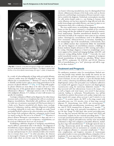
• Fig. 34.9 A thoracic computed tomography image (with contrast) from a
dog with histologically diagnosed mesothelioma. The effusion resolved after Treatment and Prognosis
2
the first of five doxorubicin chemotherapy (30 mg/m , q3wk, IV) treatments.
No satisfactory treatment exists for mesothelioma. Radical exci-
sion may benefit some animals, but usually the tumors are too
In a study of echocardiography of dogs with pericardial effusion, advanced locally and have spread by implantation early in the
a discrete cardiac mass was identified in only 5 of 15 dogs with course of disease. In one case report, a 2-year-old Siberian husky
effusion due to mesothelioma. 292 Thoracic CT may be of benefit with a solitary sclerosing mesothelioma affecting the left thoracic
to identify nodular lesions and to assess lung parenchyma in the diaphragmatic surface with pericardial and mediastinal adhesions
presence of pleural effusion 295,296 (Fig. 34.9). Dogs with neoplas- was treated with aggressive surgical resection and diaphragmatic
tic pleural effusion are more likely to be older and on CT have reconstruction using the transversus abdominis muscle. 304 The
thickening only of the parietal pleura compared with dogs with dog recovered well, but subcutaneous masses at the surgery site,
inflammatory effusion. 297 Although noted in only 3 of 20 dogs as well as hepatic and renal masses, were noted 54 days postop-
with malignant effusion, CT evidence of chest wall invasion was eratively, leading to euthanasia. Pericardiectomy may palliate
specific for neoplasia. mesothelioma patients that present with cardiac tamponade; two
Cytologic evaluation of fluid can be diagnostic for other disease dogs treated with surgery alone survived 4 and 9 months in one
processes such as infection or lymphoma but will not conclusively report. 305 In another report, the MST in five dogs treated with
diagnose mesothelioma. Mesothelial cells proliferate and exfoli- pericardiectomy was 13.6 months; three of these dogs received
ate under any circumstance associated with fluid accumulation in adjuvant IV chemotherapy (two DOX, one mitoxantrone). 306
a body cavity. Reactive mesothelial cells display many cytologic A dog treated with pericardiectomy, intrathoracic and IV cispla-
features of malignancy, making a definitive diagnosis of neoplasia tin, and IV DOX remained free of disease at 27 months. 307 In
via cytology impossible in most cases. Although one study found a report of eight dogs with pericardial mesothelioma, the MST
pericardial fluid pH analysis to be a discriminatory test to differen- was 60 days (range 15–300 days) after partial pericardiectomy.
tiate benign from malignant effusions, subsequent studies found The one dog that survived 300 days was treated with DOX and
too much overlap for the test to be of benefit. 298,299 Elevation in intracavitary cisplatin for the 4 months preceding death. 294 Tho-
pleural effusion fibronectin concentrations in dogs and cats is a racoscopic partial pericardiectomy is a less invasive procedure than
sensitive but nonspecific test for malignancy; mesothelioma can open thoracic surgical pericardiectomy and has been successfully
be ruled out if fibronectin levels are not increased. 300 performed in dogs with malignant pericardial effusions, includ-
Establishing a definitive diagnosis of malignant mesothelioma ing four dogs with mesotheliomas. 308 Although reported only in
may be difficult, particularly early in the disease. The diagnosis of one dog, thoracoscopy portal site seeding from contamination
mesothelioma requires adequate tissue sampling, preferably from with mesothelioma cells on instruments or cannulas or via leak-
an open, visually directed biopsy. Increasing availability of thora- age of malignant effusion is a potential complication. 309 Short-
coscopy and laparoscopy for small animals provides a less invasive term complications and longterm outcomes of thoracoscopic
way to evaluate these cases. 301 In either procedure, the clinician pericardial window to treat pericardial effusion, idiopathic (10) or
is encouraged to biopsy any body cavity lining and any regional neoplastic (5), were reported in 15 dogs, one of which had meso-
lymph nodes when an obvious cause for fluid accumulation is thelioma. 310 The procedure had a complication rate of 25%, low