
Page 811 - Withrow and MacEwen's Small Animal Clinical Oncology, 6th Edition
P. 811
CHAPTER 34 Miscellaneous Tumors 789
Radiographs are insensitive in the diagnosis of cardiac tumors, lesions, if seen, are most common in the areas of the right atrium
with a reported sensitivity of only 47% for cardiac HSA. 425 Many and heart base and may elevate the intrathoracic trachea. 338 Lung
metastases may also be observed.
radiographic findings are associated with the presence of pericar-
VetBooks.ir dial effusions. Animals with a large-volume pericardial effusion identifying tumors of the heart in cats and dogs. 426––428 In a study
Echocardiography is the most widely used imaging tool for
may have a globoid cardiac silhouette with crisp margins owing to
reduced cardiac motion (Fig. 34.10). Smaller fluid accumulations of 107 dogs with pericardial effusion, the sensitivity and speci-
may allow visualization of chamber contours and atrial/tumor ficity of echocardiography were 82% and 100%, respectively, for
shadows. 338 In the setting of cardiac tamponade, animals may detection of a cardiac mass; with detection of right atrial/auricular
have diminutive pulmonary arteries and veins with distention masses being slightly higher (82% sensitivity and 99% specificity)
of the caudal vena cava. In cardiac tamponade or in the setting than that of heart base tumors (74% sensitivity and 98% specific-
of obstructive mass lesions, fluid accumulations such as pleural ity). 429 The positive and negative predictive values of echocardiog-
effusion, ascites, or pulmonary edema may be observed. 423 Mass raphy were 100% and 75%, respectively, for detection of a cardiac
mass. 429 In a small study of histologically-confirmed HSA of the
right atrium and/or auricle, echocardiography had a positive pre-
dictive value of 92% (11/12) and a negative predictive value of
64% (9/14) in dogs. 428 Tumor location (extrapericardial, noncavi-
tary pericardial, and small auricular masses) and size appeared to
be the most important factors leading to false -negative results via
echocardiography. 428 This finding was supported by a larger recent
study of 51 dogs with histologically confirmed HSA of the right
atrium (Fig. 34.11a) or right auricle (Fig. 34.11b), where right
atrial tumors were more readily diagnosed (95% detection rate)
than right auricular tumors (60% detection rate). 331 Additionally,
tumor location is shown to be only moderately predictive for cor-
rectly identifying underlying tumor type (i.e., HSA vs. chemodec-
toma, etc.). 430 Pericardial effusions are commonly associated with
cardiac tumors in both cats and dogs, 385,428,429,431 being present in
42% (10 of 24) of patients with echocardiographically diagnosed
cardiac tumors in a recent study. 430 Pericardial effusion is more
commonly identified in patients with cardiac HSA, occurring in
82% of cases in a recent study. 331 Echocardiographic diagnosis
of mesothelioma is challenging, as many small lesions are below
the resolution echocardiography and a single, larger mass lesion
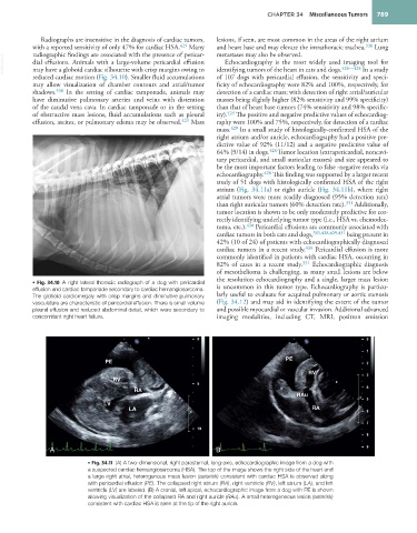
• Fig. 34.10 A right lateral thoracic radiograph of a dog with pericardial is uncommon in this tumor type. Echocardiography is particu-
effusion and cardiac tamponade secondary to cardiac hemangiosarcoma.
The globoid cardiomegaly with crisp margins and diminutive pulmonary larly useful to evaluate for acquired pulmonary or aortic stenosis
vasculature are characteristic of pericardial effusion. There is small volume (Fig. 34.12) and may aid in identifying the extent of the tumor
pleural effusion and reduced abdominal detail, which were secondary to and possible myocardial or vascular invasion. Additional advanced
concomitant right heart failure. imaging modalities, including CT, MRI, positron emission
*
PE PE
RV
RV
RA *
RAu
LV
LA RA
A B
• Fig. 34.11 (A) A two-dimensional, right parasternal, long-axis, echocardiographic image from a dog with
a suspected cardiac hemangiosarcoma (HSA). The top of the image shows the right side of the heart and
a large right atrial, heterogenous mass lesion (asterisk) consistent with cardiac HSA is observed along
with pericardial effusion (PE). The collapsed right atrium (RA), right ventricle (RV), left atrium (LA), and left
ventricle (LV) are labeled. (B) A cranial, left apical, echocardiographic image from a dog with PE is shown
allowing visualization of the collapsed RA and right auricle (RAu). A small heterogeneous lesion (asterisk)
consistent with cardiac HSA is seen at the tip of the right auricle.