
Page 812 - Withrow and MacEwen's Small Animal Clinical Oncology, 6th Edition
P. 812
790 PART IV Specific Malignancies in the Small Animal Patient
For dogs and cats with a cardiac mass and suspected neoplasia,
every effort should be made to determine the extent of disease
and the existence of primary or metastatic sites elsewhere in the
VetBooks.ir patient. In addition to echocardiography and/or other advanced
imaging modalities, a minimum database is recommended,
including a CBC, serum biochemical profile, urinalysis, coagu-
lation profile, thoracic radiographs, and abdominal ultrasound.
Concurrent splenic masses have been reported in 9 of 31 (29%)
dogs with suspected primary cardiac HSA with 42% of dogs hav-
ing metastases to other sites such as the liver, mesentery, omentum,
and lungs. 444 Conversely, this same retrospective study identified
concurrent right atrial lesion in 2 of 23 (8.7%) dogs with primary
splenic HSA whereas a previous necropsy study reported that 6
of 25 (24%) dogs with splenic HSA had a concurrent right atrial
lesion. 444,445
In a minority of cases, a diagnosis may be obtained by FNA
cytology, endomyocardial biopsy, or open surgical or thoraco-
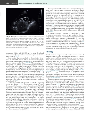
• Fig. 34.12 A right parasternal, short-axis, basilar view, echocardiographic scopic biopsy. A recent small case series of 6 dogs reported 100%
image from a dog with a suspected aortic body tumor. A large mass lesion success of obtaining a diagnostic cytology sample by FNA. This
(asterisk) consistent with an aortic body tumor is seen adjacent to the technique resulted in minor complication in two of six dogs and
cross-sectional aorta (Ao) and compressing the region of the main pul- required general anesthesia for four of six aspiration attempts. 446
monary artery branches. The right branch of the main pulmonary artery Despite this recent publication, the majority of cases remain
has extraluminal compression secondary to the heart base tumor, which treated based on anatomic location of the mass (e.g., right auricle
is creating a pressure overload on the right ventricle and subsequent right- assumed to be HSA) owing to the risks of obtaining a definitive
sided congestive heart failure in this patient.
diagnosis in the setting of limited therapeutic options.
Therapy
tomography (PET), and PET/CT, may be useful for selected
cases, particularly in preparation for possible surgical or radiation- Initial treatment for patients with cardiac tumors consists of
based therapies. 432–436 therapies directed at the secondary complications to improve
Other clinical diagnostic methods for the evaluation of car- cardiac output and hemodynamic function; however, interven-
diac or pericardial masses include pneumopericardiography, tions to manage complications such as arrhythmias and conges-
selective and nonselective angiography, gated radionuclide imag- tive heart failure will have limited efficacy unless management
ing, and endomyocardial biopsy. 338,401,437 These techniques are of the primary tumor is initiated. The use of endovascular stents
infrequently used in favor of accessible imaging by echocardiog- as palliation has been described in two dogs with cardiac masses
raphy, cardiac MRI, and CT/angiography. Cytologic evaluation obstructing venous return to the right atrium. 447 Surgical resec-
of pericardial fluid and pericardial fluid pH has been shown in tion of primary cardiac masses may be considered, but is gen-
multiple studies to be of limited utility in discriminating between erally limited to tumors arising from the right auricle (see Fig.
neoplastic and nonneoplastic causes of pericardial effusion 438–440 ; 34.11b). 330,332,448–452 Successful resection of intracardiac masses
however, in the setting of a patient with pericardial effusion and has been reported, but requires specialized anesthetic intervention
no obvious cardiac tumor by echocardiography, pericardial fluid and surgical equipment not readily available in many veterinary
cytology may offer a diagnosis in approximately 8% of cases. 441 hospitals, and preferably subspecialty training in cardiovascular
Improved diagnostic yield (20%) of pericardial fluid cytology was surgery. 351,352,365,451,453
identified in cases where the pericardial effusion had a hematocrit Subtotal pericardiectomy for dogs with heart base masses has
of less than 10%. 441 been shown to improve survival in dogs with ABTs and mesothe-
Cardiac troponin I (cTnI) appears to be useful for diagnosing lioma, regardless of whether pericardial effusion is present at the
cardiac HSA in dogs. 442,443 cTnI is a sensitive and specific marker time of diagnosis. 336,337 However, pericardiectomy alone does not
for myocardial ischemia and necrosis. Dogs with cardiac HSA had improve outcomes for dogs with cardiac HSA. 454 As dogs with
significantly higher concentrations of cTnI than did dogs with pericardial effusion treated with thoracoscopic pericardial window
idiopathic pericardial effusion. 442 In another study, the median are more likely to have recurrence of pericardial effusion than dogs
plasma cTnI concentration was higher in dogs with cardiac HSA treated with open thoracotomy and subtotal pericardiectomy, the
compared with dogs with HSA at other sites, dogs with other neo- latter is preferable for dogs expected to have prolonged survival
plasms, and dogs with pericardial effusion not caused by HSA. (i.e., ABT or mesothelioma). 455 Thoracoscopic approaches for
Furthermore, dogs with cTnI concentrations higher than 0.25 ng/ subtotal pericardiectomy and resection of cardiac masses are being
mL were likely to have cardiac HSA, and a plasma cTnI higher performed more frequently as surgeons become more experienced
than 2.45 ng/mL indicated that cardiac involvement is likely in in minimally invasive surgical techniques. 452,456,457 Resection of
dogs with confirmed HSA. 443 In practice, the measurement of right atrial or auricular masses suspected to be HSA should be
cTnI may aid in reducing the number of false-negative results by considered palliative owing to the high metastatic rate and adju-
echocardiogram. The authors routinely measure cTnI in patients vant chemotherapy is recommended to prolong survival. 332
with pericardial effusion and no obvious tumor on echocardio- DOX-based chemotherapy protocols are commonly used for
gram, using the published cutoff value of greater than 0.25 ng/mL the treatment of dogs with cardiac HSA, either as primary therapy
as a likely indication of cardiac HSA. 443 or after surgery. Protocols described for cardiac HSA include DOX