
Page 1013 - Small Animal Internal Medicine, 6th Edition
P. 1013
CHAPTER 55 Clinical Conditions of the Bitch and Queen 985
BOX 55.2 progesterone suppresses the leukocyte response to infectious
stimuli in the uterus, decreases myometrial contractility, and
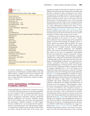
VetBooks.ir Normal Bacterial Florae of the Canine Vagina stimulates endometrial gland development and activity.
During diestrus, the nongravid uterus is flaccid and contains
Aerobic Bacteria
Pasteurella multocida endometrial gland secretions that are a growth medium for
bacteria. Bacteria reach the uterus via ascension from the
β-hemolytic Streptococci distal portion of the genitourinary tract, or less commonly
Escherichia coli
Unclassified gram - rods by hematogenous spread. Failure to clear transient bacterial
Unclassified gram + rods inhabitants from the uterus after estrus can result in pyome-
Mycoplasma tra, a septic inflammatory condition of the uterus. E. coli is
α and nonhemolytic Streptococci most commonly isolated from both bitches and queens with
Proteus pyometra (Hagman and Greko, 2005; Chen et al., 2003).
Bacillus Strong correlation exists between the onset of clinical signs
Corynebacterium of pyometra and recent heat in both species; because queens
Coagulase-positive and coagulase-negative Staphylococci are induced ovulators, their incidence may be lower.
Pseudomonas Pyometra can occur with or without purulent vulvar dis-
Klebsiella charge, depending on the patency of the cervix. Closed-
Neisseria cervix pyometra is more serious because of the potential for
Micrococcus
Haemophilus leakage of purulent fluid through the fallopian tube(s) or
Moraxella uterine rupture and resultant septic peritonitis. The classic
Acinetobacter clinical signs of pyometra include variably copious vulvar
Flavobacterium discharge, partial to complete anorexia, vomiting, lethargy,
Lactobacillus weight loss, an unkempt appearance, and polydipsia/
Enterobacter polyuria. Most pets are considered ill (lethargic, anorexic) by
Anaerobic Bacteria their owners at the time of examination. Abnormalities
Bacteroides melaninogenicus detected most frequently by physical examination include a
Corynebacterium mucopurulent to hemorrhagic vulvar discharge, a palpably
Haemophilus aphrophilus enlarged uterus, and pyrexia. Some bitches and queens have
Bacteroides no physical signs of disease other than abnormal vulvar dis-
Enterococcus
Peptostreptococcus (hemolytic and nonhemolytic) charge. Clinicopathologic evaluation most commonly dem-
Ureaplasma onstrates neutrophilic leukocytosis, hyperfibrinogenemia,
and hyperglobulinemia. Azotemia and low urinary specific
gravity can reflect nephrogenic diabetes insipidus secondary
to endotoxin elaboration by E. coli. Urine should not be
of excessive malodorous or abnormal vaginal discharge, obtained by cystocentesis if pyometra is suspected. Cytologic
vaginal mucosal inflammation, peripheral leukocytosis, and examination of the vulvar discharge shows septic inflamma-
systemic illness is significant and warrants treatment with tion (Fig. 55.39). Plasma progesterone is typically 5.0 ng/mL
antimicrobial agents. If possible, a uterine cytologic speci- or higher, typical of diestrus, although pyometra can also be
men or biopsy should be examined for evidence of inflam- initially diagnosed in early anestrus as well. Abdominal radi-
mation or infection. ography can identify a large, tubular, soft tissue density
compatible with uterine enlargement. Ultrasonography is
indicated to differentiate the enlarged fluid-filled uterus of
CYSTIC ENDOMETRIAL HYPERPLASIA/ pyometra from early pregnancy. Ultrasonographic evalua-
PYOMETRA COMPLEX tion of the uterus provides important information concern-
ing uterine wall thickness and composition (presence of
Uterine pathology (e.g., CEH) must be considered as a cause cystic structures), lumen size and content, and overall organ
of infertility in bitches and queens once all other possibilities symmetry and position. CEH is characterized by endome-
have been excluded. CEH is a hormonally dependent, pre- trial thickening with focal anechoic structures noted in the
dictable condition in the bitch that results from repeated uterine wall, representing dilated cystic glands and tortuous
cycles of progesterone stimulation inducing endometrial glandular ducts (Fig. 55.40). With advanced disease, these
glandular proliferation and secretion. Glandular changes changes do not disappear ultrasonographically during anes-
may be focal or diffuse and may interfere with implantation trus. Fluid accumulation in the uterine lumen may represent
and placentation. Definitive diagnosis of CEH requires hydrometra, mucometra, or developing pyometra and can be
biopsy at an affected site or can be confirmed by histopathol- very difficult to differentiate (echogenicity may suggest cel-
ogy post ovariohysterectomy. The CEH-pyometra complex lularity) (Figs. 55.41, A, 55.41, B, 55.42). Because of the
is a progesterone-mediated uterine disorder of both bitches potential for peritonitis, centesis of the uterus is not advo-
and queens. During the luteal phase of the estrous cycle, cated. One study found that measurement of the circulating