
Page 1014 - Small Animal Internal Medicine, 6th Edition
P. 1014
986 PART VIII Reproductive System Disorders
VetBooks.ir 0
1
2
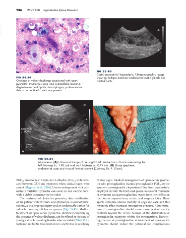
FIG 55.40
Cystic endometrial hyperplasia. Ultrasonographic image
FIG 55.39 showing multiple anechoic endometrial cystic glands and
Cytology of vulvar discharge associated with open dilated ducts.
pyometra. Numerous intra- and extracellular bacteria,
degenerative neutrophils, macrophages, proteinaceous
debris and epithelial cells are present.
0
1
2
SAGITTAL
L HORN
3
1.90cm 4
0.74cm
A B
FIG 55.41
Mucometra. (A) Ultrasound image of the sagittal left uterine horn. Cursors measuring the
full thickness (+; 1.90 cm) and wall thickness (x; 0.74 cm). (B) Gross specimen;
endometrial cysts and mucoid luminal content (Courtesy Dr. P. Olson).
PGF 2α metabolite (15-keto-13,14-dihydro-PGF 2α ) differenti- clinical signs. Medical management of open-cervix pyome-
ated between CEH and pyometra when clinical signs were tra with prostaglandins (natural prostaglandin PGF 2α or the
absent (Hagman et al., 2006). Uterine enlargement with pyo- synthetic prostaglandin cloprostenol) has been successfully
metra is variable. Pyometra can occur in one uterine horn, employed in both the bitch and queen. Successful treatment
with a viable pregnancy in the other. of pyometra using prostaglandins results from their effect on
The treatment of choice for pyometra, after stabilization the uterine myometrium, cervix, and corpora lutea. These
of the patient with IV fluids and antibiotics, is ovariohyster- agents stimulate uterine motility in dogs and cats, and this
ectomy, a challenging surgery and an undesirable option for myotonic effect increases intrauterine pressure. Administra-
valuable breeding bitches or queens (Fig. 55.43). Medical tion of prostaglandins should cause movement of uterine
treatment of open-cervix pyometra, identified clinically by contents toward the cervix because of the distribution of
the presence of vulvar discharge, can be offered in the case of prostaglandin receptors within the myometrium. Restrict-
young, valuable breeding females who are stable (Table 55.1). ing the use of prostaglandins to treatment of open-cervix
Systemic antibiotic treatment alone is ineffective in resolving pyometra should reduce the potential for complications