
Page 1041 - Small Animal Internal Medicine, 6th Edition
P. 1041
CHAPTER 57 Neonatology and Pediatrics 1013
growing healthy dogs and cats are presented in Tables 57.1 intervention before inevitable septic contamination occurs
and 57.2; the biochemical parameters from birth to approxi- may improve the prognosis in veterinary patients.
VetBooks.ir mately 8 weeks of age and up to 12 months of age in puppies weight gain, regurgitation of milk through the nares, respi-
Gastrointestinal abnormalities are suggested by poor
in Tables 57.3 and 57.4; and the biochemical parameters
from birth to approximately 8 weeks of age and up to 12
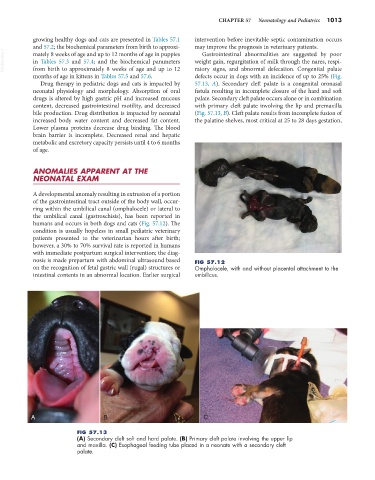
defects occur in dogs with an incidence of up to 25% (Fig.
months of age in kittens in Tables 57.5 and 57.6. ratory signs, and abnormal defecation. Congenital palate
Drug therapy in pediatric dogs and cats is impacted by 57.13, A). Secondary cleft palate is a congenital oronasal
neonatal physiology and morphology. Absorption of oral fistula resulting in incomplete closure of the hard and soft
drugs is altered by high gastric pH and increased mucous palate. Secondary cleft palate occurs alone or in combination
content, decreased gastrointestinal motility, and decreased with primary cleft palate involving the lip and premaxilla
bile production. Drug distribution is impacted by neonatal (Fig. 57.13, B). Cleft palate results from incomplete fusion of
increased body water content and decreased fat content. the palatine shelves, most critical at 25 to 28 days gestation,
Lower plasma proteins decrease drug binding. The blood
brain barrier is incomplete. Decreased renal and hepatic
metabolic and excretory capacity persists until 4 to 6 months
of age.
ANOMALIES APPARENT AT THE
NEONATAL EXAM
A developmental anomaly resulting in extrusion of a portion
of the gastrointestinal tract outside of the body wall, occur-
ring within the umbilical canal (omphalocele) or lateral to
the umbilical canal (gastroschisis), has been reported in
humans and occurs in both dogs and cats (Fig. 57.12). The
condition is usually hopeless in small pediatric veterinary
patients presented to the veterinarian hours after birth;
however, a 30% to 70% survival rate is reported in humans
with immediate postpartum surgical intervention; the diag-
nosis is made prepartum with abdominal ultrasound based FIG 57.12
on the recognition of fetal gastric wall (rugal) structures or Omphalocele, with and without placental attachment to the
intestinal contents in an abnormal location. Earlier surgical umbilicus.
A B C
FIG 57.13
(A) Secondary cleft soft and hard palate. (B) Primary cleft palate involving the upper lip
and maxilla. (C) Esophageal feeding tube placed in a neonate with a secondary cleft
palate.