
Page 1138 - Small Animal Internal Medicine, 6th Edition
P. 1138
1110 PART IX Nervous System and Neuromuscular Disorders
BOX 63.1
VetBooks.ir Vestibular Disease Clinical Findings
Central and Peripheral Vestibular Disease
Incoordination, loss of balance, disoriented
Head tilt toward lesion
Cerebellum Circling/falling/rolling toward the side of the lesion
±Ventral strabismus on side of lesion
Vomiting, salivation
8V Spontaneous or positional nystagmus (fast-phase, away
from lesion)
External
ear canal Brainstem Peripheral Vestibular Disease
Tympanic Nystagmus, when present, is horizontal or rotary
bulla No change in nystagmus direction
Postural reactions and proprioception normal
With middle/inner ear disease, may see concurrent CN7
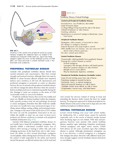
FIG 63.1 deficit and/or Horner syndrome
Anatomy of the central and peripheral vestibular system. No other cranial nerve deficits
Sensory receptors for vestibular input are located in the
membranous labyrinth of the inner ear. Input from these Central Vestibular Disease
receptors enters the brain via the vestibular portion of CN8 Occasionally indistinguishable from peripheral disease
(8V), and fibers terminate in central vestibular nuclei in the Findings that confirm disease as central:
brainstem and cerebellum. Vertical nystagmus
Nystagmus that changes direction with head position
Abnormal postural reactions on side of lesion
PERIPHERAL VESTIBULAR DISEASE Multiple cranial nerve deficits
Animals with peripheral vestibular disease should have May be depressed, stuporous, comatose
normal mentation and consciousness. They have normal
strength and postural reactions, although these tests may be Paradoxical Vestibular Syndrome (Cerebellar Lesion)
difficult to assess because affected animals have impaired Head tilt and circling away from side of lesion
balance and a tendency to fall and roll. Spontaneous and Fast-phase nystagmus toward the lesion
positional nystagmus is horizontal or rotary or alternates Horizontal, rotary, or vertical nystagmus
Abnormal postural reactions on side of lesion
between the two in animals with peripheral vestibular disease ±Multiple cranial nerve deficits on side of lesion
and will not change fast-phase direction when the animal is ±Hypermetria, truncal sway, and head tremor
held in multiple positions or examined repeatedly during the
day. Damage to inner ear receptors or the axons of CN8
occasionally results in concurrent deafness.
Peripheral vestibular lesions often involve the middle and neck toward the sternum instead of raising its head and
inner ear. Middle ear lesions localized within the tympanic extending the thoracic limbs toward the floor for weight
bulla typically produce head tilt and pathologic horizontal bearing. The diagnostic approach for bilateral peripheral ves-
or rotary nystagmus. Disorders that affect both the middle tibular disease is the same as that used in dogs and cats with
and inner ear usually have more pronounced signs of ves- unilateral peripheral vestibular disease.
tibular ataxia, falling, circling, rolling, and positional strabis-
mus. Damage to the axons of the facial nerve (CN7) within CENTRAL VESTIBULAR DISEASE
the inner ear and the sympathetic innervation to the eye Certain clinical findings only occur with central vestibu-
within the middle or inner ear can result in facial nerve lar dysfunction, but a central lesion cannot be excluded
paralysis and Horner syndrome in animals with peripheral (especially early in the course of disease) simply because
vestibular dysfunction (Fig. 63.3). these localizing signs are not evident. With time and pro-
Animals with bilateral peripheral vestibular disease do gression, however, most animals with central vestibular
not usually have a head tilt or pathologic nystagmus. They disease manifest signs indicating brainstem involvement.
typically exhibit a wide-based crouched stance, loss of Vertical nystagmus and upper motor neuron (UMN)
balance when turning or leaning to either side, and wide limb deficits are the most consistent signs that vestibular
swinging excursions of the head from side to side. Some- disease is central. Although spontaneous nystagmus can
times the ability to elicit normal physiologic nystagmus be in any direction, a vertical nystagmus or a nystagmus
(oculocephalic reflex) during head movement is lost. When that changes fast-phase direction with different head posi-
the animal is held suspended by the pelvis and lowered tions strongly suggests central vestibular disease (brainstem
toward the ground, an affected animal may curl its head and or cerebellum).