
Page 1141 - Small Animal Internal Medicine, 6th Edition
P. 1141
CHAPTER 63 Head Tilt 1113
is sedated or anesthetized for imaging, a culture should be
obtained from the external ear canal, and the ear canal and
VetBooks.ir the tympanic membrane should be carefully examined using
an otoscope or a small endoscope. If imaging suggests fluid
is present within the middle ear, a sample of that fluid should
be collected for cytologic analysis and culture. If the tym-
panic membrane is ruptured, the sample can be obtained
directly under visualization. If the tympanic membrane
appears to be intact, a myringotomy can be performed after
the external ear canal is cleansed by flushing with warm 0.9%
saline until the flush fluid obtained is clear and any excess
fluid suctioned away. Using a 22-gauge, 3.5-inch spinal
needle attached to a 6-mL syringe, the clinician punctures
the ventral tympanic membrane just caudal to the malleus
at the 6 o’clock position and gently aspirates fluid from the
middle ear into the syringe. If fluid is not obtained, 0.5 to
1 mL of sterile saline can be instilled, then aspiration can be
repeated. After the diagnostic sample is obtained, the middle A
ear should be flushed repeatedly with sterile saline to remove
exudate from the bulla.
Medical treatment of dogs and cats with bacterial OM-OI
consists of a 6- to 8-week course of systemic antibiotics, with
the choice of antibiotic based on culture and sensitivity
results. Pending culture results, antibiotic treatment can be
initiated using a broad-spectrum antibiotic such as a first-
generation cephalosporin (e.g., oral [PO] cephalexin, 22 mg/
kg q8h), a combination of amoxicillin and clavulanic acid
(Clavamox, 12.5-25 mg/kg PO q8h), or enrofloxacin (5 mg/
kg PO q12h). Identification and treatment of factors predis-
posing to otitis externa and topical or systemic antiinflam-
matory therapy are also important. If conservative treatment
does not resolve the infection or if there is radiographic B
evidence of chronic bone changes in the bulla, ventral bulla
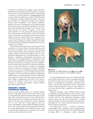
osteotomy or total ear canal ablation procedures should be FIG 63.5
performed, followed by a course of antibiotic therapy. Early Twelve-year-old Golden Retriever with (A) head and (B)
recognition of OM-OI and prompt initiation of appropriate body tilt caused by geriatric canine vestibular disease.
therapy result in a good prognosis for recovery. When facial
nerve paralysis is present, it may be permanent despite treat- Any older dog with a peracute onset of unilateral periph-
ment. Failure to aggressively treat OM-OI can result in eral vestibular disease but no other neurologic abnormali-
ascent of the infection up the nerves into the brainstem, ties should be suspected to have geriatric canine vestibular
resulting in neurologic deterioration, central vestibular disease. A careful physical examination, neurologic exami-
signs, and sometimes death. nation, and otoscopic examination should be performed.
Further extensive diagnostic testing is often delayed for
GERIATRIC CANINE a few days while the dog is supported and monitored for
VESTIBULAR DISEASE improvement.
Geriatric canine vestibular disease (i.e., old dog vestibular Diagnosis of geriatric canine vestibular disease is based
disease), an idiopathic syndrome, is the most common cause on signalment, neurologic findings, exclusion of other causes
of acute unilateral peripheral vestibular dysfunction in older of peripheral vestibular dysfunction, and alleviation of clini-
dogs, with a mean age of onset of 12.5 years. The disorder is cal signs with time. The spontaneous nystagmus usually
characterized by the very sudden onset of head tilt, loss of resolves within a few days and is replaced by a transient
balance, and ataxia with a horizontal or rotatory nystagmus positional nystagmus in the same direction. The ataxia grad-
(Fig. 63.5; see Video 58.2). Clinical signs are often very ually abates by 1 to 2 weeks, as does the head tilt. Occasion-
severe, with inability to stand, rolling and falling toward the ally the head tilt is permanent.
lesion, and vomiting. Proprioception and postural reactions The prognosis for recovery is excellent; no therapy is
are normal, although they may be difficult to assess. Facial recommended. When vomiting is severe, H 1 histaminergic
paresis and Horner syndrome are not present, and no other receptor antagonists (subcutaneous [SC] diphenhydramine,
neurologic abnormalities are observed. 2-4 mg/kg q8h), M 1 cholinergic receptor antagonists