
Page 1148 - Small Animal Internal Medicine, 6th Edition
P. 1148
1120 PART IX Nervous System and Neuromuscular Disorders
histopathology. Assessments of treatment efficacy are there- parenchymal lesions of GME, but contrast enhancement is
fore almost always based on only a presumptive diagnosis. common, reflecting inflammation. Imaging results not con-
VetBooks.ir GRANULOMATOUS clusive for neoplasia should prompt collection and analysis
of CSF.
MENINGOENCEPHALITIS
CSF analysis from dogs with GME typically reveals an
GME is an idiopathic inflammatory disorder of the CNS that increase in protein concentration and a mild to marked
occurs primarily in young adult dogs of small breeds, with mononuclear pleocytosis. Small lymphocytes, monocytes,
Poodles, toy breeds, Pekingese, and Terriers most commonly and occasional plasma cells predominate (Fig. 64.2). Ana-
affected. Large-breed dogs are occasionally affected. Most plastic mononuclear cells with abundant lacy cytoplasm are
dogs with GME are 2 to 6 years of age, although the disease sometimes present. Neutrophils are seen in two thirds of
may affect older dogs or dogs as young as 6 months. Females the samples, usually making up less than 20% of the cells. A
may be predisposed. Cats are not affected. single sample of CSF may be normal in 10% to 15% of cases.
There are three distinct forms of GME. The ocular form CSF electrophoresis typically shows evidence of blood-
is the least common and results in optic neuritis with an brain barrier disruption, and chronically affected dogs have
acute onset of blindness and dilated nonresponsive pupils dramatically increased intrathecal production of gamma
(see Chapter 61). Fundiscopic examination may reveal a globulins. Evaluation for infectious causes of meningoen-
hyperemic, edematous optic disk or hemorrhage. Dogs with cephalomyelitis through bacterial culture and appropriate
ocular GME may concurrently or later develop disseminated serum and CSF titers (Neospora, Toxoplasma, tick-borne
GME. The focal form of GME induces clinical signs sugges- diseases, West Nile virus) and antigen tests or PCR should
tive of a single enlarging space-occupying mass with neuro- always precede a presumptive diagnosis of GME. Definitive
logic signs that progress slowly over 4 to 6 months, similar diagnosis requires biopsy or necropsy for histologic exami-
to a tumor. Focal GME most often affects the caudal cranial nation, but this is rarely performed. A tentative diagnosis is
fossa, resulting in vestibulocerebellar signs or the cerebrum
causing forebrain signs including seizures. The disseminated
form of GME causes rapidly progressive signs of multifocal
or disseminated disease affecting the cerebrum, brainstem,
cerebellum, cervical spinal cord, or meninges. Affected dogs
may be febrile, and neck pain is common.
Clinical signs reflect the location and nature of the lesion.
About 20% of affected dogs exhibit seizures, circling, or
behavior change. Other common features may include brain-
stem signs such as nystagmus, head tilt, loss of balance, and
cranial nerve deficits. Cervical pain occurs in up to 10% of
dogs with GME, suggesting meningeal inflammation, focal
spinal cord involvement, or increased intracranial pressure.
Spinal cord infiltration occurs in approximately 10% of cases. A
Some dogs (50%) with the disseminated form of GME have
a fever and peripheral neutrophilia but no other evidence of
systemic disease. The disseminated form of the disease has
an acute to subacute progression over days to weeks to
months, with 25% of the cases dead within 1 week and most
dead within 21 days. The focal form is more insidious, with
slow progression over 4 to 6 months.
The diagnostic approach to patients with progressive
intracranial or spinal cord disease should include careful
systemic evaluation for infectious or neoplastic disease.
Screening laboratory tests, thoracic radiographs, abdominal
ultrasound, lymph node aspirates, and ophthalmic examina-
tion should all be performed. If these tests are unremark-
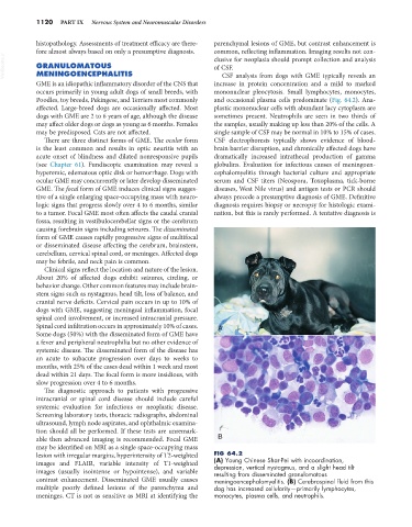
able then advanced imaging is recommended. Focal GME B
may be identified on MRI as a single space-occupying mass
lesion with irregular margins, hyperintensity of T2-weighted FIG 64.2
images and FLAIR, variable intensity of T1-weighted (A) Young Chinese Shar-Pei with incoordination,
depression, vertical nystagmus, and a slight head tilt
images (usually isointense or hypointense), and variable resulting from disseminated granulomatous
contrast enhancement. Disseminated GME usually causes meningoencephalomyelitis. (B) Cerebrospinal fluid from this
multiple poorly defined lesions of the parenchyma and dog has increased cellularity—primarily lymphocytes,
meninges. CT is not as sensitive as MRI at identifying the monocytes, plasma cells, and neutrophils.