Page 1152 - Small Animal Internal Medicine, 6th Edition
P. 1152
1124   PART IX   Nervous System and Neuromuscular Disorders


                                                                 spinal cord compression by an epidural abscess, surgery may
                                                                 be required.
  VetBooks.ir                                                    are common, particularly if the underlying source of the
                                                                   The response to antibiotic therapy is variable, and relapses
                                                                 bacterial infection cannot be resolved. The prognosis should
                                                                 be considered guarded in most cases, because even with
                                                                 appropriate therapy many animals die. An exception may be
                                                                 otogenic intracranial infections in dogs and cats, where a
                                                                 good success rate after treatment with surgical drainage and
                                                                 antibiotics has been reported.
                                                                 CANINE DISTEMPER VIRUS

             A                                                   Canine distemper virus (CDV) is a paramyxovirus that
                                                                 affects the CNS of dogs. Widespread vaccination has sub-
                                                                 stantially decreased the incidence of clinically apparent CDV
                                                                 infections in many regions, but outbreaks still occur among
                                                                 unvaccinated dogs and sporadically in vaccinated dogs.
                                                                 Clinical signs vary depending on virulence of the virus
                                                                 strain, environmental conditions, and host age and immune
                                                                 status. Most CDV infections are probably subclinical or
                                                                 are associated with mild signs of upper respiratory tract
                                                                 infection that resolve without therapy. Young, immuno-
                                                                 compromised, and unvaccinated dogs are most likely to
                                                                 develop severe generalized distemper.
                                                                   Progressive generalized infection with CDV most com-
             B                                                   monly affects unvaccinated puppies between 12 and 16
                                                                 weeks of age. The first sign of infection is a mild serous to
                                                                 mucopurulent ocular and nasal discharge followed by a dry
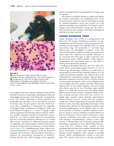
            FIG 64.3
            (A) This 4-year-old Cocker Spaniel with a chronic    cough and sometimes tonsillitis. The cough becomes moist
            retrobulbar abscess developed fever and severe depression.   and productive as pneumonia develops. Affected dogs are
            (B) Cerebrospinal fluid from the dog revealed septic   depressed, inappetent, and often febrile. Diarrhea develops
            inflammation. Postmortem examination confirmed       in some puppies and may be mild or severe. Hyperkeratosis
            communication between the retrobulbar abscess and central   of the footpads and nose, pustular dermatitis affecting the
            nervous system.                                      unhaired skin on the ventral abdomen, and severe moist
                                                                 otitis externa may also be seen. Neurologic signs typically
                                                                 begin 1 to 3 weeks after recovery from the initial systemic
            gram-negative infections, and metronidazole can be used for   illness and may include forebrain, vestibulocerebellar, brain-
            anaerobic infections. Doxycycline, trimethoprim-sulfa, and   stem, or spinal cord signs. Some of the most common neu-
            chloramphenicol may also reach therapeutic concentrations   rologic signs reported include dementia, disorientation,
            in the CNS. While inflammation persists, ampicillin and   altered mentation, seizures, circling, head tilt, nystagmus,
            amoxicillin with clavulanic acid are also effective and may   cranial nerve deficits, hypermetria, tetraparesis, and ataxia.
            be the best choice for gram-positive infections. Initial treat-  Seizures can be of any type, depending on the region of the
            ment with a combination of intravenous (IV) ampicillin   forebrain that is affected, but “chewing gum” seizures caused
            (22 mg/kg IV q6h), enrofloxacin (2.5 mg/kg IV q12h), and   by polioencephalomalacia of the temporal lobes are com-
            metronidazole (15 mg/kg IV × 1, then 7.5 mg/kg IV q8h)   monly described. Myoclonus, a repetitive rhythmic contrac-
            may be warranted if the infectious agent is unknown. When-  tion of a group of muscles resulting in repetitive flexion of a
            ever possible, antibiotics should be administered intrave-  limb or contractions of the muscles of mastication, is often
            nously for 3 to 5 days to achieve high CSF concentrations,   referred to as distemper chorea and is very common in dogs
            and then oral therapy should be continued for 4 to 8 weeks   with distemper encephalomyelitis. Anterior uveitis, optic
            after recovery. Concurrent IV fluids and systemic support   neuritis, or chorioretinitis may be detected during an oph-
            are important, and anticonvulsants should be administered   thalmologic examination in some infected dogs. Dogs sur-
            to patients having seizures (see discussion of status epilepti-  viving mild CDV infection before eruption of their
            cus in Chapter 62). Antiinflammatory drugs or glucocorti-  permanent teeth will often have irregular dental surfaces and
            coids (dexamethasone, 0.2 mg/kg IV q12h) are sometimes   brown discoloration of their teeth subsequent to virus-
            administered for the first 2 days of antibiotic treatment to   induced  enamel  hypoplasia.  Older  animals  occasionally
            minimize  the  inflammatory  consequences  of  antibiotic-  develop chronic encephalomyelitis months to years after
            induced bacterial lysis. When imaging reveals significant   prior CDV infection and recovery (old dog encephalitis),

