
Page 1189 - Small Animal Internal Medicine, 6th Edition
P. 1189
CHAPTER 66 Disorders of Peripheral Nerves and the Neuromuscular Junction 1161
VetBooks.ir
A
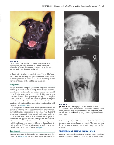
FIG 66.6
Contraction of the muscles on the left side of the face
developed in an adult dog with a 2-month history of
idiopathic left-sided facial nerve paralysis. Note the erect
left ear and nasal deviation to the left.
and cats with facial nerve paralysis caused by middle/inner
ear disease also develop peripheral vestibular signs and/or
Horner syndrome because of the close proximity of the
nerves in the area of the middle and inner ear.
Diagnosis
Idiopathic facial nerve paralysis can be diagnosed only after
excluding all other causes. A complete neurologic examina-
tion should be performed to ensure there are no other cranial
nerve deficits, ataxia, or proprioceptive deficits suggesting a
brainstem lesion. Clinicopathologic testing (i.e., complete
blood count [CBC], serum biochemistry profile, urinalysis)
is required to evaluate for systemic or metabolic disease. A B
suspicion of hypothyroidism warrants evaluation of thyroid
function (see Chapter 48). FIG 66.7
(A and B) Skull radiographs of a 4-year-old Cocker
All dogs and cats with facial nerve paralysis should be Spaniel with bilateral otitis media resulting in bilateral facial
evaluated carefully for disease of the middle and inner ear. nerve paralysis. Both bullae chambers are opacified, and
Careful otoscopic examination is important even if general the left bulla is thickened by irregular and slightly indistinct
anesthesia is required. Most animals with otitis media or new bone.
otitis interna have obvious otitis externa and a tympanic
membrane that appears abnormal or ruptured, but occasion-
ally the otoscopic examination is normal. If the suspicion for facial nerve paralysis. If keratoconjunctivitis sicca is present,
middle and inner ear disease is high, general anesthesia for the eye should be medicated as needed. The paralysis may
radiographs or CT and myringotomy to collect a sample be permanent, or spontaneous recovery may occur in 2 to
from the middle ear are warranted (Fig. 66.7). 6 weeks.
Treatment TRIGEMINAL NERVE PARALYSIS
Medical treatment for bacterial otitis media/interna is dis- Bilateral motor paralysis of the trigeminal nerves results in
cussed in Chapter 63. No treatment exists for idiopathic sudden onset of an inability to close the jaw or prehend food.