
Page 1187 - Small Animal Internal Medicine, 6th Edition
P. 1187
CHAPTER 66 Disorders of Peripheral Nerves and the Neuromuscular Junction 1159
VetBooks.ir
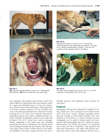
A
FIG 66.2
Mapping the region of sensory loss is important in
localizing lesions and monitoring improvement. This dog
has a caudal brachial plexus avulsion, so he has lost
superficial limb sensation distal to the elbow.
B
FIG 66.1 FIG 66.3
(A) Traumatic brachial plexus avulsion in a Chesapeake Dramatic muscle atrophy and sensory loss in a cat with
Bay Retriever. (B) Horner syndrome in the same dog. lymphoma involving the L6-S1 nerve roots.
(root signature). The insidious onset of these tumors may clinically apparent until significant spinal invasion has
make it difficult to differentiate them from lameness caused taken place.
by a vague musculoskeletal injury or nerve root compres-
sion caused by intervertebral disk disease. With progression Diagnosis
of the tumor, atrophy, weakness, and loss of reflexes may Radiographs of the spine are indicated if a neoplasm involv-
occur as the affected peripheral nerve is destroyed. Tumors ing a spinal nerve root is suspected. Nerve sheath tumors
involving the T1-T3 nerve roots commonly interrupt the rarely cause bony changes, although expanding tumors
sympathetic pathway and result in ipsilateral Horner syn- that pass through an intervertebral foramen may cause
drome. Similarly, the ipsilateral cutaneous trunci reflex will widening of the foramen as a result of pressure necrosis.
be absent if the C8-T1 ventral nerve roots are damaged. Myelography can be useful to identify spinal cord com-
Tumors originating in the spinal canal and extending pression. Electromyography and nerve conduction velocity
peripherally and tumors originating in the brachial plexus determinations may confirm the presence of a peripheral
and extending proximally into the vertebral canal will often nerve lesion and aid in localization. Deep palpation and
cause upper motor neuron (UMN) deficits in the ipsilat- ultrasound examination of the axilla under general anes-
eral hindlimb as the tumor expands, but this may not be thesia may reveal a mass. MR imaging is the best way to