
Page 1182 - Small Animal Internal Medicine, 6th Edition
P. 1182
1154 PART IX Nervous System and Neuromuscular Disorders
of spinal cord dysfunction. The enzyme deficiency itself or imaging. Instability with significant luxation can be recog-
accumulation of the metabolic intermediates within cells nized on a lateral view as widening of the space between the
VetBooks.ir causes a gradual progression of neurologic signs. Spinal dorsal lamina of the atlas and the dorsal spinous process of
the axis and dorsal displacement of the body of the axis (Fig.
signs are usually UMN in nature, although peripheral nerve
dysfunction may occur. Cortical signs (e.g., seizures) and
radiographs should be repeated with the head gently flexed
cerebellar signs (e.g., hypermetria) are more common. 65.24). If preliminary radiographs are not diagnostic, the
Signs are gradually progressive and usually obvious within to demonstrate instability.
the first year or two of life. Metabolic storage diseases are
diagnosed on the basis of the typical clinical course and Treatment
signalment; the lack of any other identifiable etiology; Emergency treatment for acute severe tetraparesis caused by
and, in some cases, organomegaly, abnormal appearance, atlantoaxial luxation should include medical treatment as for
blindness, and other readily identifiable clinical abnormali- acute spinal cord trauma (see Fig. 65.4). Medical and surgical
ties resulting from accumulation of metabolic products in treatment options have been described. Nonsurgical treat-
extraneural sites. ment should include application of a ventrally reinforced
neck brace to hold the head and neck in extension for 4 to 8
Atlantoaxial Instability and Luxation weeks, strict cage rest, and administration of analgesics. The
Normally, the atlas (C1) and axis (C2) are bound together by aim is to stabilize the neck while the ligamentous structures
ligaments. The dens, a bony projection from the cranial heal. Medical treatment has been recommended for dogs
aspect of the body of the axis, is held firmly against the floor younger than 6 months of age, those with mild neurologic
of the atlas by the transverse ligament, maintaining align- deficits, those with an acute onset of clinical signs, small dogs
ment of these two vertebrae and integrity of the spinal canal. that fracture a normal atlantoaxial articulation, and those
Malformation or absence of the dens leading to instability owners with serious financial constraints. Surgical treatment
can be seen as a congenital defect in many small breeds of is more effective but may be associated with high periopera-
dogs, including the Yorkshire Terrier, Miniature or Toy tive morbidity and mortality. Dorsal and ventral techniques
Poodle, Chihuahua, Pomeranian, Maltese, and Pekingese. are described in the Suggested Readings.
The malformation and resultant atlantoaxial instability can
lead to dorsal displacement of the axis in relation to the atlas, Prognosis
with subsequent cervical spinal cord compression and repet- In dogs with congenital atlantoaxial instability that survive
itive spinal cord trauma. Trauma may cause C1/C2 luxation, the perioperative period, the prognosis for recovery is good.
precipitating a sudden onset of cervical pain, tetraparesis, Positive outcomes are more likely if onset of signs begins
paralysis, or death. before the patient is 2 years old, signs have been present for
less than 10 months, and surgical reduction is good.
Clinical Features
Dogs with congenital atlantoaxial instability can present
with acute or chronic signs of a C1-C5 myelopathy. Signs
typically develop before 2 years of age. Clinical signs can
include neck pain (50%-75%), low head carriage, ataxia, tet-
raparesis, and postural reaction, and proprioceptive deficits
with normal to increased muscle tone and myotactic reflexes
in all four limbs. Paralysis is rare, but if it does occur it may
be accompanied by caudal brainstem signs such as hypoven-
tilation and vestibular signs. Atlantoaxial luxation secondary
to malformation should be suspected in any young (i.e., 6- to
18-month-old) toy-breed dog with a history of cervical pain,
UMN tetraparesis, or tetraplegia, whether or not there is a
history of trauma.
Diagnosis
Atlantoaxial subluxation can be diagnosed from survey
radiographs of the cervical spine, but care must be taken to
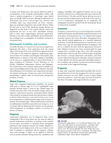
prevent inadvertent overflexion or twisting of the unstable FIG 65.24
cervical spine. Recumbent or standing lateral radiographs Atlantoaxial subluxation in a 7-month-old Bichon Frisé. The
performed with minimal restraint are usually adequate to dens rises well above its normal position, consistent with
rupture of its ligament and compression of the cervical
demonstrate dorsal displacement of the axis relative to the spinal cord. The space between the arch of the atlas and
atlas. If sedation or anesthesia will be required, a Robert the spinous process of the axis is increased. This dog had a
Jones–style bandage or splint can be applied to hold the chronic history of intermittent cervical pain and severe
neck in partial extension during induction, intubation, and ambulatory upper motor neuron tetraparesis.