
Page 1183 - Small Animal Internal Medicine, 6th Edition
P. 1183
CHAPTER 65 Disorders of the Spinal Cord 1155
Syringomyelia/Hydromyelia Ventricular dilation
Cystic accumulations of fluid within the spinal cord causing Supraoccipital bone
VetBooks.ir compression of adjacent parenchyma are being recognized Chiari malformation
with increasing frequency as advanced diagnostic imaging
techniques (i.e., CT, MRI) are used for neurologic diagnosis.
Syringomyelia is development of a CSF-filled cavity (syrinx)
within the parenchyma of the spinal cord, and hydromyelia *
is accumulation of excessive CSF within a dilated central
canal. These disorders can develop as a result of altered Basioccipital bone
CSF pressures within the spinal canal, a loss of spinal cord
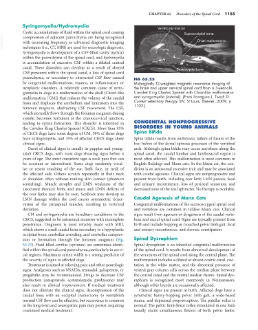
parenchyma, or secondary to obstructed CSF flow caused FIG 65.25
by congenital malformations, trauma, or inflammatory or Midsagitally T2-weighted magnetic resonance imaging of
neoplastic disorders. A relatively common cause of syrin- the brain and upper cervical spinal cord from a 3-year-old
gomyelia in dogs is a malformation of the skull (Chiari-like Cavalier King Charles Spaniel with Chiari-like malformation
malformation [CM]) that reduces the volume of the caudal and syringomyelia (asterisk). (From Bonagura J, Twedt D:
fossa and displaces the cerebellum and brainstem into the Current veterinary therapy XIV, St Louis, Elsevier, 2009, p
1102.)
foramen magnum, obstructing CSF movement. The CSF,
which normally flows through the foramen magnum during
systole, becomes turbulent at the craniocervical junction,
leading to syrinx formation. This disorder is inherited in CONGENITAL NONPROGRESSIVE
the Cavalier King Charles Spaniel (CKCS). More than 95% DISORDERS IN YOUNG ANIMALS
of CKCS dogs have some degree of CM, 50% of those dogs Spina Bifida
have syringomyelia, and 35% of affected CKCS dogs show Spina bifida results from embryonic failure of fusion of the
clinical signs. two halves of the dorsal spinous processes of the vertebral
Onset of clinical signs is usually in puppies and young- arch. Although spina bifida may occur anywhere along the
adult CKCS dogs, with most dogs showing signs before 4 spinal canal, the caudal lumbar and lumbosacral region is
years of age. The most consistent sign is neck pain that can most often affected. This malformation is most common in
be constant or intermittent. Some dogs randomly vocal- English Bulldogs and Manx cats. In the Manx cat, the con-
ize or resent touching of the ear, limb, face, or neck of dition is an autosomal recessive trait and may be associated
the affected side. Others scratch repeatedly at their neck with caudal agenesis. Clinical signs are nonprogressive and
or shoulder, often without making skin contact (phantom present from birth, including rear limb LMN paresis, fecal
scratching). Muscle atrophy and LMN weakness of the and urinary incontinence, loss of perineal sensation, and
associated thoracic limb, and ataxia and UMN deficits of decreased tone of the anal sphincter. No therapy is available.
the rear limbs may also be seen. Scoliosis may develop as
LMN damage within the cord causes asymmetric dener- Caudal Agenesis of Manx Cats
vation of the paraspinal muscles, resulting in vertebral Congenital malformations of the sacrococcygeal spinal cord
deviation. and vertebrae are common in tailless Manx cats. Clinical
CM and syringomyelia are hereditary conditions in the signs result from agenesis or dysgenesis of the caudal verte-
CKCS, suggested to be autosomal recessive with incomplete brae and sacral spinal cord. Signs are typically present from
penetrance. Diagnosis is most reliably made with MRI, birth and include hopping or crouched pelvic limb gait, fecal
which shows a small caudal fossa secondary to a hypoplastic and urinary incontinence, and chronic constipation.
occipital bone, cerebellar crowding, and cerebellar compres-
sion or herniation through the foramen magnum (Fig. Spinal Dysraphism
65.25). Fluid-filled cavities (syrinxes) are sometimes identi- Spinal dysraphism is an inherited congenital malformation
fied within the spinal cord parenchyma, particularly in cervi- of the spinal cord. It results from abnormal development of
cal regions. Maximum syrinx width is a strong predictor of the structures of the spinal cord along the central plane. The
the severity of signs in affected dogs. malformation includes a dilated or absent central canal, cavi-
Treatment is aimed at relieving pain and other neurologic tation in the white matter, and the abnormal presence of
signs. Analgesics such as NSAIDs, tramadol, gabapentin, or ventral gray column cells across the median plane between
pregabalin may be recommended. Drugs to decrease CSF the central canal and the ventral median fissure. Spinal dys-
production (omeprazole, acetazolamide, prednisone) may raphism is recognized most commonly in Weimaraners,
also result in clinical improvement. If medical treatment although other breeds are occasionally affected.
does not alleviate the clinical signs, decompression of the Clinical signs are present at birth. Affected dogs have a
caudal fossa with an occipital craniectomy to reestablish symmetric bunny-hopping pelvic limb gait, a wide-based
normal CSF flow can be effective, but recurrence is common stance, and depressed proprioception. The patellar reflex is
in the long term and neuropathic pain may persist, requiring normal. The pelvic limb flexor reflex stimulated in one limb
continued medical treatment. usually elicits simultaneous flexion of both pelvic limbs.