
Page 1180 - Small Animal Internal Medicine, 6th Edition
P. 1180
1152 PART IX Nervous System and Neuromuscular Disorders
or dorsolateral spinal cord compression that becomes clini-
cally evident when they are between 1 and 4 years of age.
VetBooks.ir Degenerative changes of the articular facets, synovial cysts,
and vertebral canal stenosis may all occur. Disk-associated
wobbler syndrome (DAWS) causes ventral compression of
the caudal cervical spinal cord in mature large-breed dogs,
especially 6- to 8-year-old Doberman Pinschers. Affected
Dobermans typically have a smaller than normal vertebral
canal, hypertrophy of the ligamentum flavum, and protru-
sion of one or more intervertebral disks leading to their signs C7
of spinal cord compression. C6
Clinical Features A
A slowly progressive course of paresis and an uncoordinated
or wobbling gait, particularly in the pelvic limbs, are charac-
teristic of CSM. Affected dogs have a broad-based rear limb
stance, ataxia, and abnormal postural reactions in the rear
limbs (which are invariably more severely affected than the
forelimbs). Neurologic findings in the forelimbs vary depend-
ing on whether spinal cord compression is centered in the
cranial cervical region or in the caudal cervical region. Dogs
with C1-C5 compression often have a floating or overreach-
ing front limb gait. Dogs with caudal cervical lesions may
have a short-strided, weak front limb gait with a weak with-
drawal reflex and pronounced atrophy of the supraspinatus C6 C7
and infraspinatus muscles over the scapula. Lameness and B
muscle atrophy in one thoracic limb or pain when traction
is applied to a limb (i.e., root signature; see Fig. 65.6) suggests
that nerve root compression is present. Slowly progressive
deterioration in neurologic status is common, but occasion- Traction
ally a traumatic episode or an acute disk extrusion results in
sudden tetraplegia. Resistance to dorsal extension of the cer-
vical spine is common, but overt cervical pain is the primary
complaint in less than 10% of dogs with CSM.
Diagnosis
The diagnosis is suspected on the basis of signalment, history,
and clinical findings. Survey radiographs are useful to rule C6 C7
out other disorders associated with cervical spinal cord com-
pression but are not definitive for CSM. Severe articular facet
changes or vertebral body malformations should raise the C
index of suspicion for CSM in a large-breed dog. Until
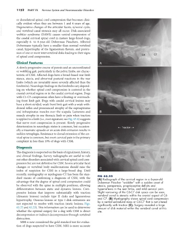
recently, myelography or myelogram-CT has been the stan- FIG 65.22
dard means of confirming a diagnosis of CSM, with the (A) Radiograph of the cervical region in a 6-year-old
advantage that the degree of spinal cord compression could Doberman Pinscher “wobbler” with a sudden onset of
be observed with the spine in multiple positions, allowing ataxia, paraparesis, proprioceptive deficits and
differentiation between static and dynamic lesions. Com- hyperreflexia in the rear limbs, and mild cervical pain.
pressive lesions that improve substantially with traction Slight narrowing of the C6-C7 disk space can be seen;
(dynamic lesions) include type II disks and ligamentous vertebral canal is stenotic within the cranial aspect of C6
hypertrophy. Osseous lesions or type I disk extrusions are and C7. (B) Myelography shows spinal cord compression
by a ventral extradural mass at C6-C7 that is not altered
not expected to resolve with traction (static lesions; Figs. significantly with traction (C). Surgery revealed a large
65.22 and 65.23). This information can be used to determine amount of disk material within the vertebral canal at this
whether the optimal surgery for a dog with CSM is direct site.
decompression or indirect decompression through vertebral
distraction.
MRI is now considered the gold standard test for evalua-
tion of dogs suspected to have CSM. MRI is more accurate