
Page 1177 - Small Animal Internal Medicine, 6th Edition
P. 1177
CHAPTER 65 Disorders of the Spinal Cord 1149
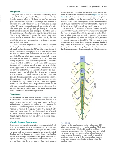
Diagnosis considerable distance within the vertebral canal caudal to the
A diagnosis of DM should be suspected in any large-breed point of termination of the spinal cord (Fig. 65.19; see also
VetBooks.ir dog with slowly progressive UMN paresis in the rear limbs. Table 65.2). This collection of nerve roots descending in the
vertebral canal is termed the cauda equina. The spinal nerves
Rear limb ataxia, a long-strided gait, toe scuffing, abnormal
postural reactions (especially knuckling), and normal to
junction, so compressive disorders affecting this region are
increased rear limb reflexes are the most common findings. from the sacral and caudal segments overlie the lumbosacral
Affected dogs are systemically normal, with no site of localiz- likely to involve the L7, sacral, and caudal nerves.
able spinal pain. Neurologic findings distinguish DM from Compression of the nerves of the cauda equina (cauda
lumbosacral disease and from orthopedic disorders such as equina syndrome, degenerative lumbosacral stenosis) is usually
hip dysplasia and bilateral anterior cruciate ligament rupture. the result of acquired type II disk protrusion at the L7/S1
The primary differential diagnoses for chronic progressive intervertebral space, together with progressive proliferation
UMN paresis in the rear limbs include DM, spinal cord of joint capsules and ligaments in the region, perhaps caused
neoplasia, spinal cord compression by articular cysts, and by excessive motion or instability. This disorder is most
type II disk protrusion. common in large-breed dogs, including German Shepherd
The antemortem diagnosis of DM is one of exclusion. dogs, Labrador Retrievers, and Belgian Malinois, and par-
Radiographs of the spine are normal, as is CSF analysis, ticularly affects male working dogs older than 5 years of age.
although a slight increase in CSF protein concentration is Rarely, compression of the cauda equina at this site could be
occasionally found. Myelography or MRI must be performed
to rule out spinal cord compression or focal spinal cord
neoplasia. Normal spinal radiographs, a cytologically normal
CSF, and normal spinal cord imaging in an older dog with L2
slowly progressive UMN signs to the pelvic limbs warrant a
diagnosis of DM. A DNA test based on the SOD1 mutation
is commercially available but will only determine which dogs L3
(homozygotes) are at risk of developing DM and which dogs L3
are carriers for the trait. The test will not identify the cause
of paraparesis in an individual dog. Recent reports suggest L4
that measuring increased concentrations of a structural L4 L5
protein of myelinated motor axons (phosphorylated neuro-
filament heavy: pNF-H) in the CSF may be useful as a bio- L6
marker for the diagnosis of DM in dogs. Definitive diagnosis L7
of DM can only be confirmed by postmortem identification L5 S1
of typical changes including axonal degeneration, demyelin- S2
S3
ation, and astroglial proliferation in the lateral funiculus and
dorsal columns of the thoracic spinal cord.
L6
Treatment
No treatment has been proven effective in dogs with DM.
Corticosteroids should not be administered, because they L7
cause muscle wasting and exacerbate muscle weakness.
Other immunosuppressive agents have not been shown to be
beneficial. Some investigators have advocated vitamins (i.e.,
vitamin E, vitamin B complex, vitamin C), omega-3 fatty S
acids, and antioxidants, but conclusive evidence of benefit
for any of these treatments is lacking. Exercise and intensive
targeted physiotherapy may be helpful in slowing disease
progression.
Cauda Equina Syndrome FIG 65.19
In dogs, the last three lumbar spinal cord segments (L5, L6, Anatomy of the cauda equina region in the dog. L5-L7
L7) are within the fourth lumbar vertebra, the sacral seg- spinal cord segments sit within the L4 vertebra. S1-S3 spinal
ments (S1, S2, S3) are within the body of the fifth lumbar cord segments are within the L5 vertebra, and the
vertebra, and the coccygeal segments are within the sixth coccygeal segments are within L6. Nerve roots from all of
the lumbar, sacral, and coccygeal spinal cord segments
lumbar vertebra. Because nerve roots from these lumbar, leave the canal through the intervertebral foramen just
sacral, and coccygeal segments of the spinal cord exit the caudal to the vertebra with the same number, so these
spinal canal through the intervertebral foramina caudal to nerve roots course a considerable distance within the
the vertebrae with the same number, they must course a vertebral canal.