
Page 1174 - Small Animal Internal Medicine, 6th Edition
P. 1174
1146 PART IX Nervous System and Neuromuscular Disorders
and abdominal ultrasound. In breeds at high risk for hem- entire axial and appendicular skeleton should be surveyed
angiosarcoma, cardiac ultrasound may also be warranted. for lytic lesions if clinical findings make multiple myeloma
VetBooks.ir Aspiration of the lymph nodes, spleen, and/or liver and likely. Soft tissue tumors of the spinal cord are almost never
visible using survey radiographs. Although myelography is a
examination of peripheral blood or bone marrow smears
may yield the diagnosis in dogs with lymphoma. Patients
cord tumors, it is relatively invasive and provides less useful
with multiple myeloma often secrete paraproteins, causing a fairly reliable method to localize and characterize spinal
hyperproteinemia and a monoclonal gammopathy. Most cats diagnostic information than MRI. CSF analysis should
with spinal lymphoma are FeLV-positive (>80%), and many always precede myelography. With tumors compressing
have obvious systemic disease and hematologic evidence of the spinal cord, CSF analysis typically reveals nonspecific
bone marrow involvement. changes, including slight increases in protein concentration
Survey radiographs of the affected region of the spine are and a mild mononuclear pleocytosis. Neoplastic cells are
recommended. Osteolysis or bone proliferation may be seen rarely identified except in cats and dogs with lymphoma
with vertebral tumors (Fig. 65.16). Fine-needle aspiration of (Fig. 65.17).
a bone lesion sometimes yields a cytologic diagnosis. The Myelography or MRI allows most spinal cord tumors
to be characterized as intramedullary, extramedullary-
intradural, or extradural. MRI adds valuable information
regarding precise tumor location and degree of spinal cord
involvement, which may be important when considering
surgical treatment and/or radiation therapy.
Treatment
Surgical decompression and attempts at complete tumor
excision are usually limited to well-encapsulated extradu-
T4 ral tumors as a referral procedure. Feline meningiomas
T3 may have a good prognosis after surgical excision. Intra-
T2 medullary tumors cannot usually be treated successfully
with surgery because of their intimate involvement with
neural tissue.
Radiation therapy may be of some benefit in dogs and
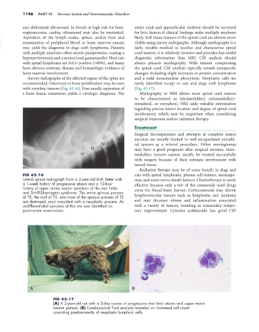
FIG 65.16 cats with spinal lymphoma, plasma cell tumors, meningio-
Lateral spinal radiograph from a 2-year-old Irish Setter with mas, and some nerve sheath tumors. Chemotherapy is rarely
a 1-week history of progressive ataxia and a 12-hour effective because only a few of the commonly used drugs
history of upper motor neuron paralysis of the rear limbs cross the blood-brain barrier. Corticosteroids may shrink
and Schiff-Sherrington syndrome. The entire spinous process
of T3, the roof of T3, and most of the spinous process of T2 lymphoreticular tumors such as lymphoma and myeloma
are destroyed, most consistent with a neoplastic process. An and may decrease edema and inflammation associated
undifferentiated sarcoma at this site was identified on with a variety of tumors, resulting in remarkable tempo-
postmortem examination. rary improvement. Cytosine arabinoside has good CSF
A B
FIG 65.17
(A) A 2-year-old cat with a 5-day course of progressive rear limb ataxia and upper motor
neuron paresis. (B) Cerebrospinal fluid analysis revealed an increased cell count
consisting predominantly of neoplastic lymphoid cells.