
Page 1171 - Small Animal Internal Medicine, 6th Edition
P. 1171
CHAPTER 65 Disorders of the Spinal Cord 1143
diagnostic testing is often necessary to identify an etiology (see Diagnosis
Chapter 64). The diagnosis of diskospondylitis is suspected after physical
VetBooks.ir Noninfectious Inflammatory Disease the affected vertebrae. Radiographic changes characteristi-
examination and confirmed by radiographic examination of
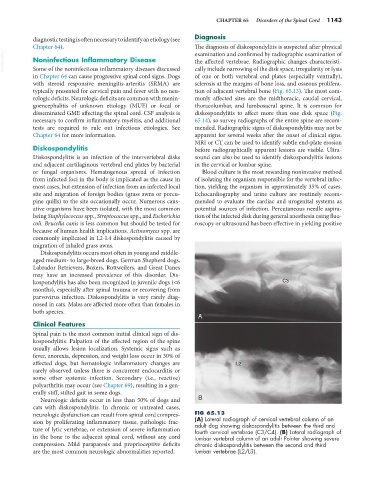
cally include narrowing of the disk space, irregularity or lysis
Some of the noninfectious inflammatory diseases discussed
in Chapter 64 can cause progressive spinal cord signs. Dogs of one or both vertebral end plates (especially ventrally),
with steroid responsive meningitis-arteritis (SRMA) are sclerosis at the margins of bone loss, and osseous prolifera-
typically presented for cervical pain and fever with no neu- tion of adjacent vertebral bone (Fig. 65.13). The most com-
rologic deficits. Neurologic deficits are common with menin- monly affected sites are the midthoracic, caudal cervical,
goencephalitis of unknown etiology (MUE) or focal or thoracolumbar, and lumbosacral spine. It is common for
disseminated GME affecting the spinal cord. CSF analysis is diskospondylitis to affect more than one disk space (Fig.
necessary to confirm inflammatory myelitis, and additional 65.14), so survey radiographs of the entire spine are recom-
tests are required to rule out infectious etiologies. See mended. Radiographic signs of diskospondylitis may not be
Chapter 64 for more information. apparent for several weeks after the onset of clinical signs.
MRI or CT can be used to identify subtle end-plate erosion
Diskospondylitis before radiographically apparent lesions are visible. Ultra-
Diskospondylitis is an infection of the intervertebral disks sound can also be used to identify diskospondylitis lesions
and adjacent cartilaginous vertebral end plates by bacterial in the cervical or lumbar spine.
or fungal organisms. Hematogenous spread of infection Blood culture is the most rewarding noninvasive method
from infected foci in the body is implicated as the cause in of isolating the organism responsible for the vertebral infec-
most cases, but extension of infection from an infected local tion, yielding the organism in approximately 35% of cases.
site and migration of foreign bodies (grass awns or porcu- Echocardiography and urine culture are routinely recom-
pine quills) to the site occasionally occur. Numerous caus- mended to evaluate the cardiac and urogenital systems as
ative organisms have been isolated, with the most common potential sources of infection. Percutaneous needle aspira-
being Staphylococcus spp., Streptococcus spp., and Escherichia tion of the infected disk during general anesthesia using fluo-
coli. Brucella canis is less common but should be tested for roscopy or ultrasound has been effective in yielding positive
because of human health implications. Actinomyces spp. are
commonly implicated in L2-L4 diskospondylitis caused by
migration of inhaled grass awns.
Diskospondylitis occurs most often in young and middle-
aged medium- to large-breed dogs. German Shepherd dogs,
Labrador Retrievers, Boxers, Rottweilers, and Great Danes
may have an increased prevalence of this disorder. Dis-
kospondylitis has also been recognized in juvenile dogs (<6 C3
months), especially after spinal trauma or recovering from
parvovirus infection. Diskospondylitis is very rarely diag-
nosed in cats. Males are affected more often than females in
both species.
A
Clinical Features
Spinal pain is the most common initial clinical sign of dis-
kospondylitis. Palpation of the affected region of the spine
usually allows lesion localization. Systemic signs such as
fever, anorexia, depression, and weight loss occur in 30% of
affected dogs, but hematologic inflammatory changes are L2
rarely observed unless there is concurrent endocarditis or
some other systemic infection. Secondary (i.e., reactive)
polyarthritis may occur (see Chapter 69), resulting in a gen-
erally stiff, stilted gait in some dogs.
Neurologic deficits occur in less than 50% of dogs and B
cats with diskospondylitis. In chronic or untreated cases,
neurologic dysfunction can result from spinal cord compres- FIG 65.13
sion by proliferating inflammatory tissue, pathologic frac- (A) Lateral radiograph of cervical vertebral column of an
adult dog showing diskospondylitis between the third and
ture of lytic vertebrae, or extension of severe inflammation fourth cervical vertebrae (C3/C4). (B) Lateral radiograph of
in the bone to the adjacent spinal cord, without any cord lumbar vertebral column of an adult Pointer showing severe
compression. Mild paraparesis and proprioceptive deficits chronic diskospondylitis between the second and third
are the most common neurologic abnormalities reported. lumbar vertebrae (L2/L3).