
Page 1167 - Small Animal Internal Medicine, 6th Edition
P. 1167
CHAPTER 65 Disorders of the Spinal Cord 1139
evaluated frequently for deterioration in neurologic status.
After 4 weeks of strict crate confinement, 3 weeks of house
VetBooks.ir confinement with no jumping or running and leash exercise
only should be recommended, followed by a gradual increase
in monitored exercise and (if necessary) a weight reduction
program. Food and water dishes should be elevated and a
chest harness should be used instead of a collar in dogs
recovering from cervical disk extrusion.
Cervical Disk Extrusion
Approximately 15% of dogs with IVD extrusion have a cervi-
cal disk, with the cranial cervical sites (especially C2-3) most
frequently affected in small-breed dogs. Dogs with a first
episode of acute neck pain and no neurologic deficits are
usually managed conservatively with strict cage confinement
and analgesics. Most dogs respond at least transiently to
conservative medical management, but a few will have
intractable pain or recurrent episodes of pain days or weeks
later. Dogs with cervical pain that does not resolve with 1 or
2 weeks of conservative management, dogs with severe pain
that cannot be controlled short term, dogs with recurrent
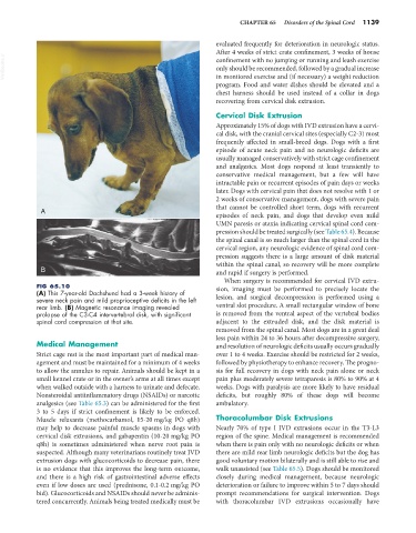
A
episodes of neck pain, and dogs that develop even mild
UMN paresis or ataxia indicating cervical spinal cord com-
pression should be treated surgically (see Table 65.4). Because
the spinal canal is so much larger than the spinal cord in the
cervical region, any neurologic evidence of spinal cord com-
pression suggests there is a large amount of disk material
within the spinal canal, so recovery will be more complete
B and rapid if surgery is performed.
When surgery is recommended for cervical IVD extru-
FIG 65.10 sion, imaging must be performed to precisely locate the
(A) This 7-year-old Dachshund had a 3-week history of
severe neck pain and mild proprioceptive deficits in the left lesion, and surgical decompression is performed using a
rear limb. (B) Magnetic resonance imaging revealed ventral slot procedure. A small rectangular window of bone
prolapse of the C3-C4 intervertebral disk, with significant is removed from the ventral aspect of the vertebral bodies
spinal cord compression at that site. adjacent to the extruded disk, and the disk material is
removed from the spinal canal. Most dogs are in a great deal
less pain within 24 to 36 hours after decompressive surgery,
Medical Management and resolution of neurologic deficits usually occurs gradually
Strict cage rest is the most important part of medical man- over 1 to 4 weeks. Exercise should be restricted for 2 weeks,
agement and must be maintained for a minimum of 4 weeks followed by physiotherapy to enhance recovery. The progno-
to allow the annulus to repair. Animals should be kept in a sis for full recovery in dogs with neck pain alone or neck
small kennel crate or in the owner’s arms at all times except pain plus moderately severe tetraparesis is 80% to 90% at 4
when walked outside with a harness to urinate and defecate. weeks. Dogs with paralysis are more likely to have residual
Nonsteroidal antiinflammatory drugs (NSAIDs) or narcotic deficits, but roughly 80% of these dogs will become
analgesics (see Table 65.3) can be administered for the first ambulatory.
3 to 5 days if strict confinement is likely to be enforced.
Muscle relaxants (methocarbamol, 15-20 mg/kg PO q8h) Thoracolumbar Disk Extrusions
may help to decrease painful muscle spasms in dogs with Nearly 70% of type I IVD extrusions occur in the T3-L3
cervical disk extrusions, and gabapentin (10-20 mg/kg PO region of the spine. Medical management is recommended
q8h) is sometimes administered when nerve root pain is when there is pain only with no neurologic deficits or when
suspected. Although many veterinarians routinely treat IVD there are mild rear limb neurologic deficits but the dog has
extrusion dogs with glucocorticoids to decrease pain, there good voluntary motion bilaterally and is still able to rise and
is no evidence that this improves the long-term outcome, walk unassisted (see Table 65.5). Dogs should be monitored
and there is a high risk of gastrointestinal adverse effects closely during medical management, because neurologic
even if low doses are used (prednisone, 0.1-0.2 mg/kg PO deterioration or failure to improve within 5 to 7 days should
bid). Glucocorticoids and NSAIDs should never be adminis- prompt recommendations for surgical intervention. Dogs
tered concurrently. Animals being treated medically must be with thoracolumbar IVD extrusions occasionally have