
Page 1164 - Small Animal Internal Medicine, 6th Edition
P. 1164
1136 PART IX Nervous System and Neuromuscular Disorders
of disk injury is most common in small-breed dogs like the
Dachshund, Toy Poodle, Pekingese, Beagle, Welsh Corgi,
VetBooks.ir Lhasa Apso, Shih Tzu, Chihuahua, and Cocker Spaniel, with
a peak incidence between 3 and 6 years of age. Dachshunds
are the breed most often presented for thoracolumbar IVD
extrusions, whereas cervical IVD extrusions are most
common in Beagles. Acute type I IVD extrusions also occa-
sionally occur in middle-aged and older large-breed dogs,
particularly in Basset Hounds, Labrador Retrievers, Dalma-
tians, Shar Peis, Border Collies, Rottweilers, Doberman Pin-
schers with caudal cervical spondylomyelopathy (CSM), and
German Shepherd dogs. Intervertebral disk extrusion is a
rare cause of clinically evident spinal cord compression in
the cat, occurring in older cats (mean age, 9.8 years) and
typically affecting the lower thoracic and lumbar regions
(most commonly, L4/L5).
Clinical Features
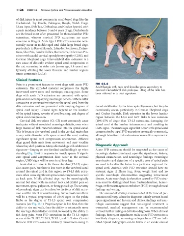
Pain is a prominent feature in most dogs with acute IVD FIG 65.6
extrusion. The extruded material compresses the highly Adult Beagle with neck and shoulder pain secondary to
innervated nerve roots and meninges, causing pain. Some cervical intervertebral disk prolapse. Lifting of the limb has
been referred to as root signature.
dogs with acute IVD extrusion are presented with spinal
pain and no accompanying neurologic deficits. Others suffer
concussive or compressive injury to the spinal cord from the
disk extrusion and are presented with varying degrees of dorsal stabilization by the intercapital ligaments, but they do
spinal cord injury. Clinical signs depend on the location occasionally occur, particularly in German Shepherd dogs
of the spinal injury, severity of cord bruising, and degree of and Cocker Spaniels. Disk extrusion in the lower lumbar
spinal cord compression. region between the L3/4 and L6/7 disks is less common
Cervical disk extrusions (C1-C5) most commonly cause (10%-15% of dogs) than T3-L3 extrusions, damaging the
neck pain without associated neurologic deficits, even when spinal cord at the lumbar intumescence and resulting in
large masses of disk material extrude into the spinal canal. LMN signs. The neurologic signs that occur with spinal cord
This is because the vertebral canal in the cervical region has compression by type I IVD extrusions are usually symmetric,
a very wide diameter with space around the cord, making although lateralized disk extrusions can result in asymmetric
significant spinal cord compression uncommon. Affected signs.
dogs guard their neck from movement and may vocalize
when they shift position. Many affected dogs will exhibit root Diagnostic Approach
signature—limping on one forelimb and holding it up when Acute IVD extrusion should be suspected as the cause of
standing (Fig. 65.6) in response to muscle spasm. If signifi- neurologic dysfunction based upon the signalment, history,
cant spinal cord compression does occur in the cervical physical examination, and neurologic findings. Neurologic
region, UMN signs will be seen in all four legs. examination and detection of a specific area of spinal pain
Acute disk extrusion in the thoracolumbar (T3-L3) region are used to localize the lesion to a particular region of the
also causes pain, but there is less room in the vertebral canal spinal cord. Animals with IVD extrusion should have no
around the spinal cord in this region, so T3-L3 disk extru- systemic signs of illness (e.g., fever, weight loss) and no
sions often cause significant spinal cord compression as well specific neurologic abnormalities suggesting intracranial
as back pain. Mildly affected dogs may be neurologically disease. Acute neurologic dysfunction caused by IVD extru-
normal but stand with an arched back and exhibit pain on sion must be distinguished from fracture/luxation, hemor-
movement, spinal palpation, or being picked up. The severity rhage, or fibrocartilaginous embolism (FCE) through clinical
of neurologic signs can be related to the force of disk extru- findings and testing.
sion and the extent of cord bruising, but in most cases there The amount of workup recommended at the time of pre-
will be a typical progression of UMN signs in the pelvic sentation will vary. When the diagnosis is fairly certain based
limbs as the degree of T3-L3 spinal cord compression upon signalment and history, and clinical findings and neu-
worsens (see Fig. 65.1). Proprioception is lost first, then the rologic assessment suggest that nonsurgical treatment is
ability to rise and walk, then the ability to voluntarily move warranted, medical management can be recommended
the rear legs, then bladder control, followed by the ability to without further testing or definitive diagnosis. When clinical
feel deep pain. Most IVD extrusions in the T3-L3 region findings, history, or signalment make acute IVD extrusion a
occur at the T11/12, T12/13, T13/L1, and L1/2 sites. Cranial less likely diagnosis, screening radiographs or CT are indi-
thoracic IVD extrusions are relatively uncommon owing to cated. Spinal radiographs can be taken in an awake animal