
Page 1166 - Small Animal Internal Medicine, 6th Edition
P. 1166
1138 PART IX Nervous System and Neuromuscular Disorders
VetBooks.ir T12
T13 T12
T12 T13
L1
L1 T13
A
L1
T12 T13
L1
D B C
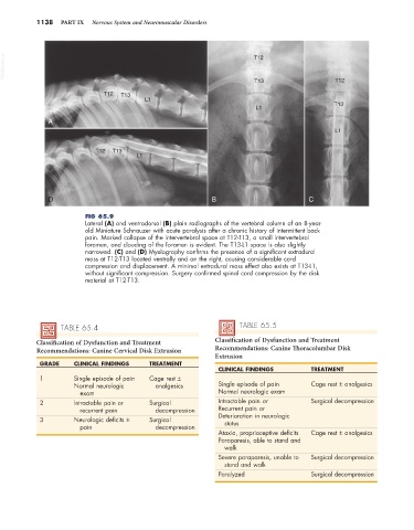
FIG 65.9
Lateral (A) and ventrodorsal (B) plain radiographs of the vertebral column of an 8-year-
old Miniature Schnauzer with acute paralysis after a chronic history of intermittent back
pain. Marked collapse of the intervertebral space at T12-T13, a small intervertebral
foramen, and clouding of the foramen is evident. The T13-L1 space is also slightly
narrowed. (C) and (D) Myelography confirms the presence of a significant extradural
mass at T12-T13 located ventrally and on the right, causing considerable cord
compression and displacement. A minimal extradural mass effect also exists at T13-L1,
without significant compression. Surgery confirmed spinal cord compression by the disk
material at T12-T13.
TABLE 65.4 TABLE 65.5
Classification of Dysfunction and Treatment Classification of Dysfunction and Treatment
Recommendations: Canine Cervical Disk Extrusion Recommendations: Canine Thoracolumbar Disk
Extrusion
GRADE CLINICAL FINDINGS TREATMENT
CLINICAL FINDINGS TREATMENT
1 Single episode of pain Cage rest ±
Normal neurologic analgesics Single episode of pain Cage rest ± analgesics
exam Normal neurologic exam
2 Intractable pain or Surgical Intractable pain or Surgical decompression
recurrent pain decompression Recurrent pain or
3 Neurologic deficits ± Surgical Deterioration in neurologic
status
pain decompression
Ataxia, proprioceptive deficits Cage rest ± analgesics
Paraparesis, able to stand and
walk
Severe paraparesis, unable to Surgical decompression
stand and walk
Paralyzed Surgical decompression