
Page 1170 - Small Animal Internal Medicine, 6th Edition
P. 1170
1142 PART IX Nervous System and Neuromuscular Disorders
by damage at the brachial or lumbosacral intumescence
(C6-T2 or L4-S3) are less likely to fully recover. Animals that
VetBooks.ir have lost the ability to perceive painful stimuli in the affected
limbs are unlikely to recover the ability to ambulate without
assistance.
ATLANTOAXIAL INSTABILITY
Although some dogs with congenital atlantoaxial instability
will be presented acutely for neck pain and tetraparesis,
many affected dogs have slowly progressive waxing and
waning tetraparesis due to repeated cervical spinal cord
injury. This condition will be discussed with progressive
spinal cord disorders affecting young animals. Traumatic
fracture of the dens leading to subluxation can occur in any
dog or cat and will result in acute UMN dysfunction in all
limbs. Management should be as described for acute spinal
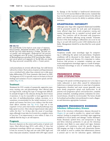
FIG 65.12 trauma.
This adult Border Collie had an acute onset of lameness,
flaccid paresis, decreased sensation, and hyporeflexia in NEOPLASIA
the left rear limb while retrieving a Frisbee. The limb was
not painful, and radiographs, cerebrospinal fluid analysis, Neoplasms usually cause neurologic signs by compress-
and myelogram were all normal. A presumptive diagnosis ing or infiltrating the spinal cord parenchyma. Neoplastic
of fibrocartilaginous embolism (FCE) involving the lumbar conditions will be discussed in this chapter with chronic
and sacral spinal cord segments on the left side was made. progressive spinal cord diseases. It is important to realize,
This dog recovered uneventfully within a 3-week period. however, that primary or metastatic neoplasia can cause
acute nonprogressive neurologic signs as a result of intrapa-
renchymal hemorrhage or lysis of vertebral bones, leading
cord parenchyma in severely affected dogs, but mild lesions to fracture.
will not be evident. The location of spinal cord hyperintensity
over a vertebral body rather than centered over a disk space
helps differentiate FCE from traumatic disk based on MRI. PROGRESSIVE SPINAL
The diagnosis of FCE is typically made on the basis of clinical CORD DYSFUNCTION
findings and exclusion of acute compressive spinal cord dis-
orders (Fig. 65.12). Damage to the spinal cord that progresses over a few days to
weeks (subacute) is most often caused by inflammatory
Treatment (infectious or immune) processes or some type of neoplasia.
Treatment for FCE consists of nonspecific supportive mea- Degenerative disorders and most cancers generally cause
sures, nursing care, and physiotherapy. Most affected dogs more slowly progressive spinal cord dysfunction. In all
are large breeds, making this type of management difficult. patients with progressive spinal cord dysfunction, complete
In animals brought to the clinician during the first 6 hours patient evaluation, including systemic evaluation for extra-
of paralysis, it may be reasonable to treat aggressively with neural disease, should be recommended. The lesion should
one dose of methylprednisolone sodium succinate, as is be localized and ancillary tests performed to reach a diagno-
sometimes recommended for the initial treatment of acute sis and determine appropriate treatment.
spinal cord trauma, but there is no evidence that this treat-
ment affects outcome (see Fig. 65.4). Cage rest is not
required—in fact, early intervention with physiotherapy may SUBACUTE PROGRESSIVE DISORDERS
help speed recovery. Most clinical improvement takes place Infectious Inflammatory Disease
within the first 7 to 10 days after the onset of neurologic Most of the infectious inflammatory diseases discussed in
signs, although it may take 6 to 8 weeks for a complete return Chapter 64 can result in myelitis (i.e., spinal cord inflam-
to function. If no improvement is seen within 21 days, it is mation), leading to progressive neurologic signs suggesting
unlikely that the dog or cat will improve. multifocal or focal spinal cord damage. Canine distemper,
Rocky Mountain spotted fever, and neosporosis in dogs
Prognosis and feline infectious peritonitis in cats are the infectious
Recovery depends on the extent and location of spinal cord disorders most likely to cause spinal cord signs. Systemic
injury. Most affected dogs (85%) walk within 21 days, but evaluation will sometimes reveal the specific diagnosis.
permanent neurologic deficits are common. The prognosis MR imaging can help to localize and characterize lesions.
is best for recovery in dogs and cats with UMN signs and CSF analysis is used to confirm that inflammatory disease
intact deep pain sensation. Animals with LMN signs caused is present and to test for infectious agents. Additional