
Page 1165 - Small Animal Internal Medicine, 6th Edition
P. 1165
CHAPTER 65 Disorders of the Spinal Cord 1137
to look for characteristic features of disk disease and to rule disk extrusion and can be very difficult to diagnose once CSF
out other diseases (e.g., diskospondylitis, lytic vertebral has been altered by instilling myelographic contrast material
VetBooks.ir tumor, fracture, atlantoaxial luxation). into the subarachnoid space (see discussion of myelography,
Chapter 59).
Radiographic observation of calcified disks confirms the
CT can be used as an adjunct to myelography or as the
presence of generalized IVDD, but, unless there is dorsal
displacement of mineralized disk material into the spinal sole diagnostic procedure to demonstrate spinal cord com-
canal, this does not necessarily implicate the disk extrusion pression by an extruded disk and to eliminate other bone-
as the cause of neurologic dysfunction. Radiographic changes related reasons for spinal cord signs (fracture, luxation,
consistent with extrusion of a disk in the thoracolumbar vertebral lysis). CT is very quick, can often be performed
region include a narrowed or wedged disk space, a small or under sedation instead of general anesthesia, and has diag-
cloudy intervertebral foramen (“horse’s head”), narrowing of nostic accuracy similar to myelography for diagnosis and
the facetal joints, and a calcified density in the spinal canal localization of calcified extruded disks. CT myelography
above the involved disk (Figs. 65.7 and 65.8). Many dogs may be necessary to determine lateralization of IVD hernia-
with disk extrusion have multiple abnormal sites, however, tion when the extruded disk is not calcified or severe spinal
and radiographs cannot determine which is the active site cord swelling is present.
causing the current problem. Myelography or advanced MRI is the best diagnostic method for localizing the
diagnostic imaging (i.e., CT, MRI) will be required to defini- site and the side of extruded disks with nearly 100% accu-
tively localize the site of an extruded disk causing spinal cord racy (Fig. 65.10). MRI also allows evaluation of the cord
compression in animals in which surgical treatment is being parenchyma for injury and edema, which may be associ-
considered. ated with prognosis for recovery in patients with loss of
Myelography was once the standard imaging modality deep pain sensation. However, MRI is slower than CT, less
for diagnosing and localizing acute disk extrusion in dogs, readily available, and more expensive, requiring general
but this has largely been replaced with the less invasive anesthesia.
and more diagnostic CT and MRI (Fig. 65.9). Myelogra-
phy is a good test to demonstrate the site of disk extrusion Treatment Recommendations
but does not (without concurrent CT) reliably determine Treatment recommendations for dogs with acute IVD extru-
whether more of the disk material is located on the left or sion are based on location of the spinal cord injury and
right side of the cord—important information for surgical severity of signs noted at the time of presentation (Tables
planning. Collection and analysis of CSF should always be 65.4 and 65.5). Treatment options are conservative (medical)
recommended before proceeding with a myelogram, because and surgical. Surgery should be recommended when decom-
inflammatory CNS disorders (granulomatous meningoen- pression will significantly increase the likelihood, speed, and
cephalitis [GME], others) can be clinically very similar to completeness of recovery.
C6
T13 L1
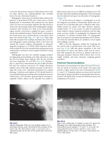
FIG 65.8
FIG 65.7 Lateral plain radiograph of vertebral column of a 4-year-old
Lateral radiograph of the cervical vertebral column of an Pekingese with acute intervertebral disk prolapse. The
adult dog showing acute intervertebral disk prolapse at the intervertebral space between T13 and L1 is narrowed, the
C6-C7 site. The intervertebral space is narrowed, and a intervertebral foramen (“horse’s head”) is small, and a
calcified density can be seen in the spinal canal above the calcified density can be seen in the spinal canal above the
disk space. T13-L1 disk space.