
Page 1173 - Small Animal Internal Medicine, 6th Edition
P. 1173
CHAPTER 65 Disorders of the Spinal Cord 1145
CHRONIC PROGRESSIVE DISORDERS
Neoplasia
VetBooks.ir Tumors that grow and compress or infiltrate spinal cord
parenchyma frequently cause chronic, progressively worsen-
ing signs of spinal cord dysfunction. Spinal tumors can be
primary or metastatic. The most common tumors affecting
the spinal cord in the dog are extradural tumors arising from
the vertebral body (e.g., osteosarcoma, chondrosarcoma,
fibrosarcoma, myeloma) and extradural soft tissue tumors,
including metastatic hemangiosarcoma, carcinoma, liposar-
coma, and lymphoma. Intradural extramedullary tumors
such as meningiomas, neuroepithelioma, and peripheral
nerve sheath tumors are also common, accounting for 35% A
of all spinal tumors. Intramedullary tumors are relatively
rare in the dog, with the exception of metastatic hemangio-
sarcoma. Lymphoma can be extradural, intradural/
extramedullary, or intramedullary in the dog and is usually
a manifestation of multicentric disease. Lymphoma is the
only common spinal tumor in the cat, and in 85% of cats
with spinal lymphoma the tumor is also found in extraneural
sites.
Most spinal cord tumors occur in middle-aged and older
dogs, with the mean age at the time of diagnosis being 5 to
6 years. Two noteworthy exceptions are lymphoma (which
can affect dogs of any age) and neuroepithelioma, a primary
intradural extramedullary tumor that has a predilection for
T10-L1 in young dogs, particularly German Shepherd dogs B
and Golden Retrievers. In addition, vertebral osteomas may
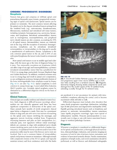
occur in young dogs and result in spinal cord compression, FIG 65.15
as can cartilaginous exostoses, benign proliferative lesions of (A) A 3-month-old Golden Retriever puppy with spinal pain
the bone indistinguishable from neoplasia except by biopsy and progressive upper motor neuron (UMN) signs in both
(Fig. 65.15; see also Fig. 59.2). Spinal lymphoma is most rear limbs resulting from a vertebral osteoma. (B) Magnetic
resonance imaging showing severe compressive spinal cord
common in young (mean age, 4 years) adult feline leukemia damage from the caudal aspect of the T4 vertebral body
(FeLV)-positive cats. Certainly, spinal neoplasia cannot be extending caudally through the T6 vertebral body.
eliminated as a differential diagnosis strictly on the basis of
signalment.
are paralyzed, it is not uncommon for animals with intra-
Clinical Features medullary neoplasms affecting the central cord to become
Clinical signs are usually insidious and related to tumor loca- incontinent while still able to walk.
tion. Early diagnosis is difficult because neurologic abnor- Differential diagnoses must include other disorders that
malities are not clinically apparent until there has been cause slowly progressive neurologic dysfunction, including
significant compression or destruction of the spinal cord. type II disk protrusion and degenerative myelopathy (DM).
Many animals have months of slowly progressive clinical Rapidly growing extradural tumors such as lymphoma and
signs before a diagnosis is made. Pain may be a prominent primary or metastatic intramedullary tumors sometimes
feature in dogs and cats with nerve root tumors encroaching cause rapidly progressive neurologic signs more typical of
on the spinal cord, tumors involving the meninges, and inflammatory myelitis. Peracute paresis/paralysis is occa-
aggressive tumors involving vertebral bone. Progressively sionally seen in dogs or cats with tumor-associated hemor-
worsening lameness and pain on limb manipulation (i.e., rhage or vertebral pathologic fractures.
radicular pain, root signature) without initial neurologic
deficits are common in dogs with peripheral nerve sheath Diagnosis
tumors involving nerve roots in the cervical or lumbar intu- Whenever a neoplasm is considered as a differential diagno-
mescence. An ipsilateral Horner syndrome and/or loss of the sis for spinal cord dysfunction, a thorough physical examina-
panniculus reflex may be seen if the thoracic nerve roots are tion, clinicopathologic evaluation, and systemic imaging are
involved. Pain is not a common feature of intramedullary necessary to look for sites of primary tumor, metastases, and
spinal cord primary tumors or metastases. Although animals evidence of associated systemic disease. Fundic examination,
with compressive lesions of the T3-L3 spinal cord typically palpation of lymph nodes, and rectal examination should be
maintain urinary and fecal continence until after the limbs performed, as well as thoracic and abdominal radiographs