
Page 1175 - Small Animal Internal Medicine, 6th Edition
P. 1175
CHAPTER 65 Disorders of the Spinal Cord 1147
penetration so may also be used to treat lymphoreticular normal cytology and slightly increased protein, consistent
tumors. with a noninflammatory chronic compressive myelopathy.
VetBooks.ir Spinal Articular Cysts Treatment usually consists of spinal cord decompression,
cyst drainage, and arthrodesis of the facetal joint, and usually
Cysts arising from the joint capsule of spinal facetal joints
eration and bony proliferation of multiple thoracolumbar
can, through enlargement, cause chronic progressive focal produces excellent results. A similar syndrome with degen-
extradural compression of the spinal cord or nerve roots. articular facets causing spinal cord compression has been
These cysts can result from an outpouching of the synovium reported as a hereditary condition in 4- to 10-month-old
(i.e., synovial cysts), or they may arise from mucinous Shiloh Shepherds.
degeneration of periarticular connective tissue (i.e., gan-
glion cysts). Synovial cysts and ganglion cysts are clinically Spinal Arachnoid Diverticula (Cysts)
indistinguishable, and both arise secondary to degenerative Focal accumulations of CSF in cyst-like structures within the
changes in the facetal joints. Degenerative changes occur subarachnoid space can lead to slowly progressive, nonpain-
because of congenital malformations, vertebral instability, or ful spinal cord compression in young dogs (Fig. 65.18). The
trauma. Signs are referable to the site and degree of resulting cyst-like structures containing CSF may represent a con-
spinal cord or nerve root compression. Young giant breeds of genital intradural arachnoid diverticulum (most common)
dogs such as Mastiffs, Great Danes, and Bernese Mountain or a pocket caused by adhesions in the subarachnoid space
Dogs most commonly develop single or multiple cysts in secondary to trauma, disk extrusion or protrusion, or ver-
the cervical region, which cause a UMN myelopathy with tebral malformations. The cervical region and the caudal
proprioceptive ataxia, progressive tetraparesis, and occasion- thoracic region are most often affected, with most diver-
ally cervical pain. Synovial cysts occur in 20% of dogs with ticula in the dorsal aspect of the vertebral canal. A CSF flow
CSM (wobbler syndrome). Older dogs, particularly German disturbance may cause a functional one-way valve, so that
Shepherd dogs, have been identified with thoracolumbar or as CSF fills the diverticulum it enlarges and causes com-
lumbosacral articular cysts that cause spinal cord or cauda pression of the spinal cord. Young large-breed male dogs
equina compression. are most likely to be affected by cervical diverticula, with
Radiographs may reveal degenerative changes of the Rottweilers overrepresented. Small-breed dogs, especially
articular facets. Diagnosis of synovial cysts is best accom- Pugs and French Bulldogs have a tendency to have thoraco-
plished with MR imaging. The cysts are well-circumscribed lumbar diverticula. Cats are rarely affected. Myelography or
extradural masses associated with the articular processes on MRI reveals accumulation of CSF at the site (see Fig. 65.18).
one or both sides of the vertebral canal. CSF analysis reveals Exploration, fenestration of the diverticula with durotomy,
A B
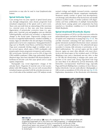
FIG 65.18
Lateral (A) and ventrodorsal (B) views of a myelogram from a 10-month-old Akita with
progressive hypermetria of all four limbs and mild paraparesis. A well-defined bulbous
dilation of the dorsal subarachnoid space communicating with the rest of the
subarachnoid space was present at C2-C3, suggesting an arachnoid diverticulum.
Surgical exploration and marsupialization resulted in rapid and persistent (>6 years) return
to normal gait.