
Page 1179 - Small Animal Internal Medicine, 6th Edition
P. 1179
CHAPTER 65 Disorders of the Spinal Cord 1151
VetBooks.ir
L7 S
A
S
L6 L7
B C
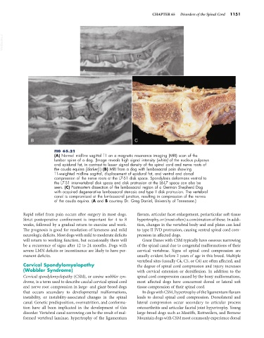
FIG 65.21
(A) Normal midline sagittal T1 on a magnetic resonance imaging (MRI) scan of the
lumbar spine of a dog. (Image reveals high signal intensity [white] of the nucleus pulposus
and epidural fat, in contrast to lesser signal density of the spinal cord and nerve roots of
the cauda equina [darker].) (B) MRI from a dog with lumbosacral pain showing
T1-weighted midline sagittal, displacement of epidural fat, and ventral and dorsal
compression of the nerve roots at the L7-S1 disk space. Spondylosis deformans ventral to
the L7-S1 intervertebral disk space and disk protrusion at the L6-L7 space can also be
seen. (C) Postmortem dissection of the lumbosacral region of a German Shepherd Dog
with acquired degenerative lumbosacral stenosis and type II disk protrusion. The vertebral
canal is compromised at the lumbosacral junction, resulting in compression of the nerves
of the cauda equina. (A and B courtesy Dr. Greg Daniel, University of Tennessee.)
Rapid relief from pain occurs after surgery in most dogs. flavum, articular facet enlargement, periarticular soft tissue
Strict postoperative confinement is important for 4 to 8 hypertrophy, or (most often) a combination of these. In addi-
weeks, followed by a gradual return to exercise and work. tion, changes in the vertebral body and end plates can lead
The prognosis is good for resolution of lameness and mild to type II IVD protrusion, causing ventral spinal cord com-
neurologic deficits. Most dogs with mild to moderate deficits pression in affected dogs.
will return to working function, but occasionally there will Great Danes with CSM typically have osseous narrowing
be a recurrence of signs after 12 to 24 months. Dogs with of the spinal canal due to congenital malformations of their
severe LMN deficits or incontinence are likely to have per- cervical vertebrae. Signs of spinal cord compression are
manent deficits. usually evident before 2 years of age in this breed. Multiple
vertebral sites (usually C4, C5, or C6) are often affected, and
Cervical Spondylomyelopathy the degree of spinal cord compression and injury increases
(Wobbler Syndrome) with cervical extension or dorsiflexion. In addition to the
Cervical spondylomyelopathy (CSM), or canine wobbler syn- spinal cord compression caused by the bony malformations,
drome, is a term used to describe caudal cervical spinal cord most affected dogs have concurrent dorsal or lateral soft
and nerve root compression in large- and giant-breed dogs tissue compression of their spinal cord.
that occurs secondary to developmental malformations, In dogs with CSM, hypertrophy of the ligamentum flavum
instability, or instability-associated changes in the spinal leads to dorsal spinal cord compression. Dorsolateral and
canal. Genetic predisposition, overnutrition, and conforma- lateral compression occur secondary to articular process
tion have all been implicated in the development of this osteoarthritis and articular facetal joint hypertrophy. Young
disorder. Vertebral canal narrowing can be the result of mal- large-breed dogs such as Mastiffs, Rottweilers, and Bernese
formed vertebral laminae, hypertrophy of the ligamentum Mountain dogs with CSM most commonly experience dorsal