
Page 1205 - Small Animal Internal Medicine, 6th Edition
P. 1205
CHAPTER 67 Disorders of Muscle 1177
ventral neck flexion, an inability to jump, and a tendency to
sit or lie down after walking short distances. Muscle pain
VetBooks.ir may be evident. Neurologic examination reveals normal
mentation, cranial nerves, proprioception, and reflexes.
Diagnosis is made on the basis of clinical features,
increases of serum CK and AST activities, and multifocal
EMG abnormalities. Many affected cats (70%) are slightly
hypokalemic, suggesting a possible relationship between this
disorder and hypokalemic polymyopathy. Because some
clinical features of PM also mimic mild thiamine deficiency,
evaluating the cat’s response to thiamine injections (intra-
muscular [IM], 10-20 mg/day) and correcting hypokalemia
are recommended before proceeding with extensive diag-
nostic testing for PM.
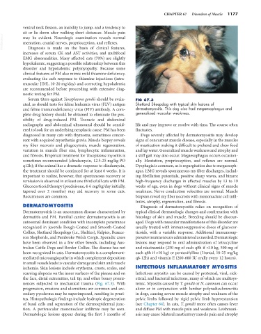
Serum titers against Toxoplasma gondii should be evalu- FIG 67.3
ated, as should tests for feline leukemia virus (FLV) antigen Shetland Sheepdog with typical skin lesions of
and feline immunodeficiency virus (FIV) antibody. A com- dermatomyositis. This dog also had megaesophagus and
plete drug history should be obtained to eliminate the pos- generalized muscular weakness.
sibility of drug-induced PM. Thoracic and abdominal
radiographs and abdominal ultrasound should be consid- life and may improve or resolve with time. The course often
ered to look for an underlying neoplastic cause. PM has been fluctuates.
diagnosed in many cats with thymoma, sometimes concur- Dogs severely affected by dermatomyositis may develop
rent with acquired myasthenia gravis. Muscle biopsy reveals signs of concurrent muscle disease, especially in the muscles
my fiber necrosis and phagocytosis, muscle regeneration, of mastication making it difficult to prehend and chew food
variation in muscle fiber size, lymphocytic inflammation, and lap water. Generalized muscle weakness and atrophy and
and fibrosis. Empirical treatment for Toxoplasma myositis is a stiff gait may also occur. Megaesophagus occurs occasion-
sometimes recommended (clindamycin, 12.5-25 mg/kg PO ally. Mentation, proprioception, and reflexes are normal.
q12h); if the animal has a dramatic response to clindamycin, Dysphagia is common, as is regurgitation due to megaesoph-
the treatment should be continued for at least 6 weeks. It is agus. EMG reveals spontaneous my fiber discharges, includ-
important to realize, however, that spontaneous recovery or ing fibrillation potentials, positive sharp waves, and bizarre
remission is observed in at least one third of all cats with PM. high-frequency discharges in affected muscles by 13 to 19
Glucocorticoid therapy (prednisone, 4-6 mg/kg/day initially, weeks of age, even in dogs without clinical signs of muscle
tapered over 2 months) may aid recovery in some cats. weakness. Nerve conduction velocities are normal. Muscle
Recurrences are common. biopsies reveal my fiber necrosis with mononuclear cell infil-
trates, atrophy, regeneration, and fibrosis.
DERMATOMYOSITIS Diagnosis of dermatomyositis relies on recognition of
Dermatomyositis is an uncommon disease characterized by typical clinical dermatologic changes and confirmation with
dermatitis and PM. Familial canine dermatomyositis is an histology of skin and muscle. Breeding should be discour-
autosomal dominant condition with incomplete penetrance aged. Dogs with muscular manifestations of this disorder are
recognized in juvenile Rough-Coated and Smooth-Coated usually treated with immunosuppressive doses of glucocor-
Collies, Shetland Sheepdogs (i.e., Shelties), Kelpies, Beauce- ticoids, with a variable response. Additional immunosup-
ron Shepherds, and Pembroke Welsh Corgis. Sporadic cases pressive treatments are administered as needed. Dermatologic
have been observed in a few other breeds, including Aus- lesions may respond to oral administration of tetracycline
tralian Cattle Dogs and Border Collies. The disease has not and niacinamide (250 mg of each q8h if <10 kg, 500 mg of
been recognized in cats. Dermatomyositis is a complement- each q8h if >10 kg) or pentoxifylline (Trental, 10-25 mg/kg
mediated microangiopathy in which complement deposition q8-12h) and vitamin E (200-600 IU orally every 12 hours).
in small vessels leads to vascular damage and skin and muscle
ischemia. Skin lesions include erythema, crusts, scales, and INFECTIOUS INFLAMMATORY MYOSITIS
scarring alopecia on the inner surfaces of the pinnae and on Infectious myositis can be caused by protozoal, viral, rick-
the face, distal extremities, tail tip, and other boney promi- ettsial, and bacterial infections, many of which are multisys-
nences subjected to mechanical trauma (Fig. 67.3). With temic. Myositis caused by T. gondii or N. caninum can occur
progression, erosions and ulcerations are common and sec- alone or in conjunction with lumbar polyradiculoneuritis
ondary pyoderma may be superimposed, resulting in pruri- in dogs, causing severe muscle atrophy and weakness of the
tus. Histopathologic findings include hydropic degeneration pelvic limbs followed by rigid pelvic limb hyperextension
of basal cells and separation of the dermoepidermal junc- (see Chapter 64). In cats, T. gondii more often causes fever
tion. A perivascular mononuclear infiltrate may be seen. and diffuse PM with muscle pain and weakness. Leishmani-
Dermatologic lesions appear during the first 3 months of asis may cause bilateral masticatory muscle pain and atrophy