
Page 1209 - Small Animal Internal Medicine, 6th Edition
P. 1209
CHAPTER 67 Disorders of Muscle 1181
occur. In Miniature Schnauzers, Australian Cattle dogs, INVOLUNTARY ALTERATIONS IN
and Jack Russell Terriers, mutant skeletal muscle chloride MUSCLE TONE AND MOVEMENT
VetBooks.ir channel alleles have been identified, and genetic testing Tetanus, opisthotonos, myoclonus, and dyskinesias are
is available.
Clinical signs include generalized muscle stiffness and
that are not the result of muscle disease. Tetanus is a sus-
hypertrophy that begin at a young age (i.e., 1-4 months) all involuntary alterations of muscle tone or movement
and difficulty rising after a period of inactivity. Dogs with tained tonic contraction of the muscles. Opisthotonos is a
myotonia are neurologically normal. No abnormalities of very severe form of tetanus in which spasm of the limb
proprioception or mentation exist. Cold weather, excite- and neck muscles results in lateral recumbency with dor-
ment, sudden changes in position or posture, and exercise siflexion of the neck and extensor rigidity of the limbs.
exacerbate the clinical signs. Affected dogs may remain in Myoclonus is the rhythmic repetitive contraction of a par-
rigid recumbency for up to 30 seconds if they are suddenly ticular group of muscles. Dyskinesias, a group of poorly
placed in lateral recumbency. Hypertrophy of the proximal defined movement disorders that can be hard to differen-
appendicular and axial skeletal muscles is common. Serum tiate from partial seizures, were previously discussed in
CK and AST activities may be mildly increased, indicating Chapter 62.
muscle fiber necrosis. Bizarre high-frequency discharges that
wax and wane (“dive-bomber sound”) are revealed by EMG OPISTHOTONOS
and, when present, confirm the diagnosis. Muscle biopsy Opisthotonos is a very severe form of sustained muscular
alone is rarely diagnostic. Membrane-stabilizing agents such contraction resulting in dorsiflexion of the neck and exten-
as procainamide (10-30 mg/kg PO q6h) and the sodium sor rigidity of the limbs. Opisthotonos can be seen during
channel blocker mexiletine (Mexitil [Boehringer Ingel- seizure activity, as a component of generalized extensor
heim], 8 mg/kg PO q8h) have been beneficial in the treat- rigidity in patients with tetanus, or in animals with decere-
ment of some cases. Avoidance of cold temperatures is also brate or decerebellate rigidity (see Fig. 58.9).
advised. Most dogs are euthanized because of the severity of
their signs. TETANUS
Tetanus, defined as sustained contraction of extensor muscles
INHERITED METABOLIC MYOPATHIES without relaxation, is most often seen in dogs and cats and
Genetically based metabolic myopathies caused by a bio- is due to infection with the bacterium Clostridium tetani and
chemical defect of the skeletal muscle energy system are its production of tetanospasmin toxin. C. tetani is a gram-
infrequently described in dogs and cats. All such disorders positive anaerobic bacillus that produces spores that persist
cause signs of muscle dysfunction, including exercise intol- for long periods in the environment. If a deep wound or an
erance, muscular weakness, a stiff stilted gait, muscle pain, area of tissue damage becomes contaminated with these
muscle tremors, and muscle atrophy. Mitochondrial myopa- spores, the spores may be anaerobically converted to a veg-
thies, glycogen storage diseases, lipid storage myopathies, etative form, and a toxin (tetanospasmin) is produced. The
and disorders causing nemaline rod accumulation within toxin ascends peripheral nerves to the spinal cord, where it
myofibers have all been reported. Genetic tests are available blocks release of neurotransmitter from the inhibitory inter-
for a few disorders. Establishing the precise cause of a meta- neurons (Renshaw cells), releasing extensor muscles from
bolic myopathy can be difficult because of the wide range of inhibition and resulting in tetany. Cats are more resistant to
biochemical abnormalities that can arise and the co- the toxin than dogs.
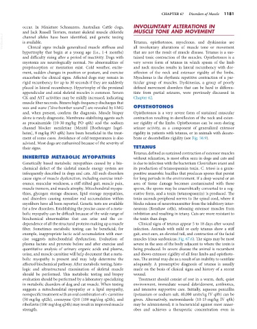
dependence of all the structural proteins making up a muscle Clinical signs of tetanus appear 5 to 18 days after wound
fiber. Sometimes metabolic testing can be beneficial; for infection. Animals with mild or early tetanus show a stiff
example, inappropriate lactic acid accumulation with exer- gait, erect ears, an elevated tail, and contraction of the facial
cise suggests mitochondrial dysfunction. Evaluation of muscles (risus sardonicus; Fig. 67.6). The signs may be most
plasma lactate and pyruvate before and after exercise and severe in the area of the body adjacent to where the toxin is
quantitative analysis of urinary organic acids and plasma, being produced. In severe disease the animal is recumbent
urine, and muscle carnitine will help document that a meta- and shows extensor rigidity of all four limbs and opisthoto-
bolic myopathy is present and may help determine the nos. The animal may die as a result of an inability to ventilate
affected biochemical pathway. After metabolic testing, histo- adequately. A presumptive diagnosis of tetanus is usually
logic and ultrastructural examination of skeletal muscle made on the basis of clinical signs and history of a recent
should be performed. This metabolic testing and biopsy wound.
evaluation should be performed by a laboratory specializing Treatment should consist of rest in a warm, dark, quiet
in metabolic disorders of dog and cat muscle. When testing environment, immediate wound debridement, antibiotics,
suggests a mitochondrial myopathy or a lipid myopathy, and intensive supportive care. Initially, aqueous penicillin
nonspecific treatment with an oral combination of L-carnitine (potassium or sodium salt, 40,000 units/kg IV q8h) can be
(50 mg/kg q12h), coenzyme Q10 (100 mg/dog q24h), and given. Alternatively, metronidazole (10-15 mg/kg IV q8h)
riboflavin (100 mg/dog q24h) may result in improved muscle may be administered; it is bactericidal against most anaer-
strength. obes and achieves a therapeutic concentration even in