
Page 151 - Small Animal Internal Medicine, 6th Edition
P. 151
CHAPTER 6 Acquired Valvular and Endocardial Disease 123
VetBooks.ir
A B
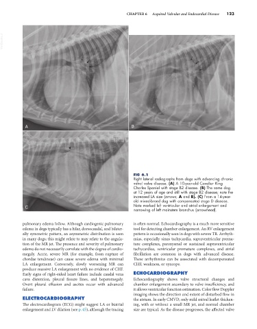
FIG 6.1
Right lateral radiographs from dogs with advancing chronic
mitral valve disease. (A) A 10-year-old Cavalier King
Charles Spaniel with stage B2 disease. (B) The same dog
at 12 years of age and still with stage B2 disease; note the
increased LA size (arrows, A and B). (C) From a 14-year-
old mixed-breed dog with compensated stage D disease.
C Note marked left ventricular and atrial enlargement and
narrowing of left mainstem bronchus (arrowhead).
pulmonary edema follow. Although cardiogenic pulmonary is often normal. Echocardiography is a much more sensitive
edema in dogs typically has a hilar, dorsocaudal, and bilater- tool for detecting chamber enlargement. An RV enlargement
ally symmetric pattern, an asymmetric distribution is seen pattern is occasionally seen in dogs with severe TR. Arrhyth-
in many dogs; this might relate to may relate to the angula- mias, especially sinus tachycardia, supraventricular prema-
tion of the MR jet. The presence and severity of pulmonary ture complexes, paroxysmal or sustained supraventricular
edema do not necessarily correlate with the degree of cardio- tachycardias, ventricular premature complexes, and atrial
megaly. Acute, severe MR (for example, from rupture of fibrillation are common in dogs with advanced disease.
chordae tendineae) can cause severe edema with minimal These arrhythmias can be associated with decompensated
LA enlargement. Conversely, slowly worsening MR can CHF, weakness, or syncope.
produce massive LA enlargement with no evidence of CHF.
Early signs of right-sided heart failure include caudal vena ECHOCARDIOGRAPHY
cava distention, pleural fissure lines, and hepatomegaly. Echocardiography shows valve structural changes and
Overt pleural effusion and ascites occur with advanced chamber enlargement secondary to valve insufficiency, and
failure. it allows ventricular function estimation. Color flow Doppler
imaging shows the direction and extent of disturbed flow in
ELECTROCARDIOGRAPHY the atrium. In early CMVD, only mild mitral leaflet thicken-
The electrocardiogram (ECG) might suggest LA or biatrial ing, with or without a small MR jet, and normal chamber
enlargement and LV dilation (see p. 45), although the tracing size are typical. As the disease progresses, the affected valve