
Page 153 - Small Animal Internal Medicine, 6th Edition
P. 153
CHAPTER 6 Acquired Valvular and Endocardial Disease 125
VetBooks.ir
A B
C
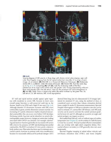
FIG 6.3
Varying degrees of MR severity in three dogs with chronic mitral valve disease, seen with
color flow Doppler imaging from the left apical 4-chamber view (the LV is at top of each
image, LA at bottom). (A) Mild MR in a 10-year-old Miniature Schnauzer. (B) Moderately
severe MR in a different, older Miniature Schnauzer. (C) Severe MR, from the same dog
as in Fig. 6.2, B. Comparing (A) with (C), note the increasing width of the flow
disturbance at its origin at the mitral valve, the greater color mixing (representing turbulent
flow) and intensity within the left atrium, and also the prominent left atrial enlargement in
(B) and (C). Note: Color is visible only within the sector outlined by the green line. LA,
Left atrium; LV, left ventricle; MR, mitral regurgitation.
LV wall and septal motion usually appear quite vigor- derived from long-axis two-dimensional (2-D) images opti-
ous with moderate to severe MR, because in most cases mized for maximal LV size, using the method of discs, is
overall pump function is well preserved until late in the considered more accurate than volume estimation derived
disease. Small to no EPSS and a high FS are seen (Fig. 6.4). from a single M-mode LV dimension measurement. Spectral
Although diastolic LV dimension increases with MR, when Doppler interrogation of MR jet acceleration rate also can be
contractility is good, systolic dimension remains normal. An used to estimate LV contractility (dP/dtmax), and MR peak
increasing LV systolic dimension implies impaired myocar- velocity can be used to estimate the systolic pressure gradi-
dial contractility; this can occur even before CHF develops. ent between LA and LV, although an eccentric jet angle and
Declining systolic function can be identified on serial echo- mitral prolapse can impair accuracy.
cardiography exams; however, changes in ventricular loading Pericardial fluid (blood), with or without signs of cardiac
associated with CMVD can interfere. For example, shorten- tamponade (see Chapter 9), can be evident if a full thickness
ing and ejection fractions generally are increased with severe LA tear has occurred. Mild pericardial effusion (transudate)
MR, even in the presence of CHF and declining myocardial also can develop with right-sided congestive failure. Peri-
contractility. The ESVI normalizes LV end systolic volume to cardial effusion secondary to CHF generally does not cause
body surface area. This index has been used to estimate myo- tamponade.
cardial systolic function in patients with valve insufficiency Spectral Doppler imaging of mitral inflow velocity and
because it is minimally influenced by preload changes. ESVI isovolumic relaxation time (IVRT), and tissue Doppler