
Page 252 - Small Animal Internal Medicine, 6th Edition
P. 252
224 PART I Cardiovascular System Disorders
corticosteroid therapy in pathologic thrombosis is unclear. heart. Hyperthyroidism could be a risk factor for ATE in cats
However, TE disease is relatively common in animals receiv- independent of its cardiac effects. A rare cause of ATE could
VetBooks.ir ing exogenous corticosteroids and in those with hyperadre- occur in cats with atrial septal defect, if a venous embolus
crossed from RA to LA. In some cases of feline ATE, no
nocorticism (see next paragraph). Other predisposing factors
usually coexist in these cases as well.
Systemic arterial emboli usually lodge at the aortic trifur-
TE disease occurs in some dogs with spontaneous hyper- predisposing condition is identified.
adrenocorticism. This endocrinopathy has been associated cation (so-called “saddle thrombus” or, more correctly,
with decreased fibrinolysis (resulting from increased PAI “saddle embolus”; Fig. 12.1), but iliac, femoral, renal, bra-
activity) and high levels of several coagulation factors. chial, and other arteries can be affected depending on
Corticosteroids result in hypercoagulable thromboelastogra- embolus size and flow path. Besides obstructing flow in the
phy (TEG) tracings in normal dogs. Diabetes mellitus is affected artery, thromboemboli release vasoactive substances
occasionally associated with TE disease in dogs. Platelet that induce vasoconstriction and compromise collateral
hyperaggregability and possibly hypofibrinolysis are thought blood flow around the obstructed vessel. Tissue ischemia
to be involved. Greyhounds appear to be predisposed to TE results and causes further damage and inflammation, includ-
disease despite lack of detectable hemostatic or cardiovascu- ing ischemia-reperfusion injury after blood flow to the area
lar abnormalities; one proposed mechanism is abnormalities is restored. An ischemic neuromyopathy occurs in the
of homocysteine metabolism (hyperhomocysteinemia). affected limb(s), with peripheral nerve dysfunction and
Occasionally, a patient with clinically relevant TE disease degeneration, as well as pathologic changes in associated
does not have any detectable abnormality that can result in muscle tissue. Coronary thromboembolism with myocardial
hypercoagulability. necrosis has occurred in cats with cardiac disease, especially
severe HCM or infective endocarditis, as well as from carci-
noma emboli.
PULMONARY THROMBOEMBOLISM
Clinical Features
Pulmonary thromboemboli (PTE) in dogs are associated Arterial TE in cats usually causes acute and dramatic clinical
with HWD, IMHA, neoplasia, DIC, sepsis, hyperadrenocor- signs secondary to tissue ischemia (Fig. 12.2). Male cats
ticism, protein-losing nephropathy, pancreatitis, trauma, appear to be at higher risk for ATE, but this gender bias
hypothyroidism, and right atrial (RA) thrombi related to appears to be related to the prevalence of HCM. Common
infection or neoplasia. physical examination findings of cats with ATE can be sum-
Pulmonary TE disease appears to be rare in cats com- marized with the mnemonic “5 Ps”: pain, pallor (pale color-
pared with dogs, except in those with HWD (see Chapter ation of affected pawpad[s]), paresis or plegia (of affected
10). Nevertheless, PTE have been associated with a variety limb[s]), pulselessness (weak or absent pulse in affected
of systemic and inflammatory disorders in cats, including limb[s]), and poikilothermia (decreased temperature of
neoplasia, HWD, anemia (probably immune-mediated), affected compared with unaffected limb[s]). Additional clin-
pancreatitis, glomerulonephritis, encephalitis, pneumonia, ical abnormalities are summarized in Box 12.2.
heart disease, sepsis, glucocorticoid administration, protein-
losing enteropathy, and hepatic lipidosis.
Pulmonary TE disease is an important cause of precapil-
lary pulmonary hypertension, and can lead to right ven-
tricular hypertrophy and even right-sided congestive heart
failure (CHF). See Chapters 19 and 25 for further informa-
tion about pulmonary thromboembolism and pulmonary
hypertension.
SYSTEMIC ARTERIAL
THROMBOEMBOLISM IN CATS
Cardiomyopathy, mainly HCM, is the most common under-
lying disease in cats with arterial thromboembolism (ATE)
(see Chapter 8). Thrombi initially form in the left heart,
usually in an enlarged LA or auricle, and can become quite
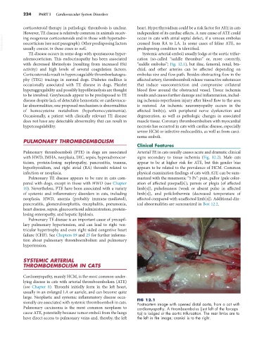
large. Neoplastic and systemic inflammatory disease occa- FIG 12.1
sionally are associated with systemic thromboemboli in cats. Postmortem image with opened distal aorta, from a cat with
Pulmonary carcinoma is the most common neoplasm to cardiomyopathy. A thromboembolus (just left of the forceps
cause ATE, potentially because tumor emboli from the lungs tip) is lodged at the aortic trifurcation. The rear limbs are to
have direct access to pulmonary veins and, thereby, the left the left in the image; cranial is to the right.