
Page 253 - Small Animal Internal Medicine, 6th Edition
P. 253
CHAPTER 12 Thromboembolic Disease 225
VetBooks.ir
A B
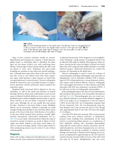
FIG 12.2
(A) Cat with thromboembolism to the distal aorta. The left rear limb was dragged behind
as the cat tried to walk; there was slightly better function in the right rear limb. (B) The
pads of the left rear paw (right side of image) in this cat were paler as well as cooler
compared with the left forepaw (left side of image).
Signs of poor systemic perfusion usually are present. on physical examination. If the diagnosis is not straightfor-
Hypothermia and azotemia are common. A heart murmur, ward, obtaining a small amount of peripheral blood from
gallop sound, or arrhythmia often is identified, but these an affected limb might be helpful. Blood glucose values are
signs are not always evident even with underlying heart lower, whereas lactate values are higher, in blood from limbs
disease. Clinical signs of heart disease before the ATE event affected by ATE compared with unaffected limbs or centrally
are absent in many cases. Tachypnea and open-mouth obtained samples. Abdominal ultrasound can confirm the
breathing are common in cats with acute arterial emboliza- presence of a thrombus in the distal aorta.
tion. Although these signs often relate to the onset of CHF, Thoracic radiography is used to screen for evidence of
they also occur in cats without overt CHF. These respira- cardiomyopathy, including cardiomegaly (and especially LA
tory signs could represent a pain response or result from enlargement), and to determine whether CHF is present.
increased pulmonary venous pressure. Thoracic radiographs Signs of CHF include pulmonary venous distension, pulmo-
should be obtained as soon as possible, because it is impor- nary edema, and/or pleural effusion. Thoracic radiographs
tant to determine whether pulmonary edema underlies the also could suggest the presence of other diseases potentially
respiratory signs. associated with ATE (e.g., pulmonary carcinoma, HWD). A
Peripheral limb functional deficits depend on the area few affected cats have no radiographic abnormalities.
embolized, as well as the extent and duration of arterial Echocardiography depicts the type of myocardial disease,
blockage. However, distal aortic embolization occurs in most and sometimes the presence of an intracardiac thrombus as
cases, so bilateral hindlimb deficits are most common. Acute well (see Fig. 8.4). Echocardiography also can reveal cavi-
hindlimb paresis without palpable femoral pulses is typical. tary effusions (pleural or pericardial effusion) suggestive of
Motor function in the rear limbs is minimal to absent in CHF. Some degree of LA enlargement is seen the majority
most cases, although the cat can usually flex and extend (>90%) of cats with ATE. An LA dimension of greater than
the hips. Sensation to the lower limbs is poor. Hindlimb 20 mm (measured from the two-dimensional, long-axis,
deficits can be asymmetric. Emboli occasionally are small four-chamber view) may increase the risk for ATE, though
enough to lodge more distally in only one limb, which causes only approximately half of cats with ATE have this degree
paresis of the lower limb alone. Embolization of an axillary of LA dilation.
or the more distal brachial artery produces (usually right) Cats with ATE often have azotemia. This can be prerenal,
forelimb monoparesis. Intermittent claudication (see pp. resulting from poor systemic perfusion or dehydration;
230–231) occurs rarely. Thromboemboli within the renal or primary renal, resulting from embolization of the renal
mesenteric arterial circulation can result in failure of these arteries or preexisting kidney disease; or a combination of
organs and death. Emboli to the brain could induce sei- both. Metabolic acidosis, DIC, electrolyte abnormalities
zures or various neurologic deficits. Clinical signs related to (especially low serum sodium, calcium, potassium, and ele-
other predisposing disease may be evident in cats without vated phosphorus), and stress hyperglycemia are common.
cardiomyopathy. Hyperkalemia can develop secondary to ischemic muscle
damage and reperfusion. Skeletal muscle damage and necro-
Diagnosis sis are accompanied by rapid elevation in CK; ALT and AST
Feline ATE usually is diagnosed clinically based on a combi- activities become elevated within 12 hours of the ATE event
nation of acute onset paraparesis and absent femoral pulses and peak by 36 hours. Myoglobinuria may also occur