
Page 485 - Small Animal Internal Medicine, 6th Edition
P. 485
CHAPTER 29 Disorders of the Oral Cavity, Pharynx, and Esophagus 457
VetBooks.ir
A B
C D
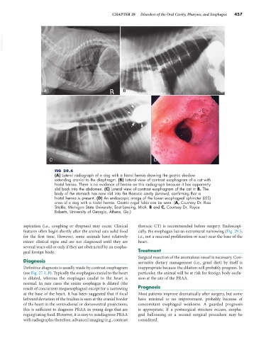
FIG 29.4
(A) Lateral radiograph of a dog with a hiatal hernia showing the gastric shadow
extending cranial to the diaphragm. (B) Lateral view of contrast esophagram of a cat with
hiatal hernia. There is no evidence of hernia on this radiograph because it has apparently
slid back into the abdomen. (C) Lateral view of contrast esophagram of the cat in B. The
body of the stomach has now slid into the thoracic cavity (arrows), confirming that a
hiatal hernia is present. (D) An endoscopic image of the lower esophageal sphincter (LES)
area of a dog with a hiatal hernia. Gastric rugal folds can be seen. (A, Courtesy Dr. Russ
Stickle, Michigan State University, East Lansing, Mich. B and C, Courtesy Dr. Royce
Roberts, University of Georgia, Athens, Ga.)
aspiration (i.e., coughing or dyspnea) may occur. Clinical thoracic CT) is recommended before surgery. Endoscopi-
features often begin shortly after the animal eats solid food cally, the esophagus has an extramural narrowing (Fig. 29.5;
for the first time. However, some animals have relatively i.e., not a mucosal proliferation or scar) near the base of the
minor clinical signs and are not diagnosed until they are heart.
several years old or only if they are obstructed by an esopha-
geal foreign body. Treatment
Surgical resection of the anomalous vessel is necessary. Con-
Diagnosis servative dietary management (i.e., gruel diet) by itself is
Definitive diagnosis is usually made by contrast esophagram inappropriate because the dilation will probably progress. In
(see Fig. 27.3, B). Typically the esophagus cranial to the heart particular, the animal will be at risk for foreign body occlu-
is dilated, whereas the esophagus caudal to the heart is sion at the site of the PRAA.
normal. In rare cases the entire esophagus is dilated (the
result of concurrent megaesophagus) except for a narrowing Prognosis
at the base of the heart. It has been suggested that if focal Most patients improve dramatically after surgery, but some
leftward deviation of the trachea is seen at the cranial border have minimal to no improvement, probably because of
of the heart in the ventrodorsal or dorsoventral projections, concomitant esophageal weakness. A guarded prognosis
this is sufficient to diagnose PRAA in young dogs that are is appropriate. If a postsurgical stricture occurs, esopha-
regurgitating food. However, it is easy to misdiagnose PRAA geal ballooning or a second surgical procedure may be
with radiographs; therefore, advanced imaging (e.g., contrast considered.