
Page 486 - Small Animal Internal Medicine, 6th Edition
P. 486
458 PART III Digestive System Disorders
VetBooks.ir
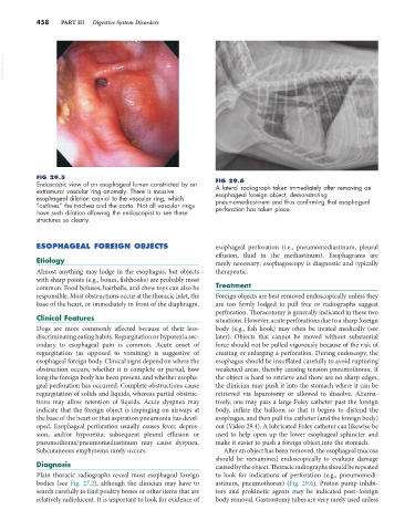
FIG 29.5 FIG 29.6
Endoscopic view of an esophageal lumen constricted by an A lateral radiograph taken immediately after removing an
extramural vascular ring anomaly. There is massive esophageal foreign object, demonstrating
esophageal dilation cranial to the vascular ring, which pneumomediastinum and thus confirming that esophageal
“outlines” the trachea and the aorta. Not all vascular rings perforation has taken place.
have such dilation allowing the endoscopist to see these
structures so clearly.
ESOPHAGEAL FOREIGN OBJECTS esophageal perforation (i.e., pneumomediastinum, pleural
effusion, fluid in the mediastinum). Esophagrams are
Etiology rarely necessary; esophagoscopy is diagnostic and typically
Almost anything may lodge in the esophagus, but objects therapeutic.
with sharp points (e.g., bones, fishhooks) are probably most
common. Food boluses, hairballs, and chew toys can also be Treatment
responsible. Most obstructions occur at the thoracic inlet, the Foreign objects are best removed endoscopically unless they
base of the heart, or immediately in front of the diaphragm. are too firmly lodged to pull free or radiographs suggest
perforation. Thoracotomy is generally indicated in these two
Clinical Features situations. However, acute perforations due to a sharp foreign
Dogs are more commonly affected because of their less- body (e.g., fish hook) may often be treated medically (see
discriminating eating habits. Regurgitation or hyporexia sec- later). Objects that cannot be moved without substantial
ondary to esophageal pain is common. Acute onset of force should not be pulled vigorously because of the risk of
regurgitation (as opposed to vomiting) is suggestive of creating or enlarging a perforation. During endoscopy, the
esophageal foreign body. Clinical signs depend on where the esophagus should be insufflated carefully to avoid rupturing
obstruction occurs, whether it is complete or partial, how weakened areas, thereby causing tension pneumothorax. If
long the foreign body has been present, and whether esopha- the object is hard to retrieve and there are no sharp edges,
geal perforation has occurred. Complete obstructions cause the clinician may push it into the stomach where it can be
regurgitation of solids and liquids, whereas partial obstruc- retrieved via laparotomy or allowed to dissolve. Alterna-
tions may allow retention of liquids. Acute dyspnea may tively, one may pass a large Foley catheter past the foreign
indicate that the foreign object is impinging on airways at body, inflate the balloon so that it begins to distend the
the base of the heart or that aspiration pneumonia has devel- esophagus, and then pull the catheter (and the foreign body)
oped. Esophageal perforation usually causes fever, depres- out (Video 29.4). A lubricated Foley catheter can likewise be
sion, and/or hyporexia; subsequent pleural effusion or used to help open up the lower esophageal sphincter and
pneumothorax/pneumomediastinum may cause dyspnea. make it easier to push a foreign object into the stomach.
Subcutaneous emphysema rarely occurs. After an object has been removed, the esophageal mucosa
should be reexamined endoscopically to evaluate damage
Diagnosis caused by the object. Thoracic radiographs should be repeated
Plain thoracic radiographs reveal most esophageal foreign to look for indications of perforation (e.g., pneumomedi-
bodies (see Fig. 27.2), although the clinician may have to astinum, pneumothorax) (Fig. 29.6). Proton pump inhibi-
search carefully to find poultry bones or other items that are tors and prokinetic agents may be indicated post–foreign
relatively radiolucent. It is important to look for evidence of body removal. Gastrostomy tubes are very rarely used unless