
Page 482 - Small Animal Internal Medicine, 6th Edition
P. 482
454 PART III Digestive System Disorders
In animals with severe aspiration, gastrostomy tubes the pharynx where it is aspirated but never expelled. In other
can be used to bypass the esophagus, and some animals cases, material is expelled but re-swallowed or re-eaten by
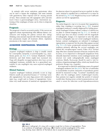
VetBooks.ir with gastrostomy tubes respond well for varying periods the animal (Fig. 29.3 A,B). Weight loss may occur if sufficient
calories are lost by regurgitation.
of time. These animals may still regurgitate saliva and also
food if there is gastroesophageal reflux. Intermittent suc-
tioning of the esophagus at home can be done in desperate Diagnosis
situations. The initial diagnostic step is to document that regurgitation
rather than vomiting is occurring (see p. 391). Acquired
Prognosis esophageal weakness is usually diagnosed by finding gener-
The prognosis is hard to accurately predict. If the owners are alized esophageal dilation without evidence of obstruction
aggressive about experimenting with different dietary con- on plain or contrast imaging (see Fig. 27.3, A). Severity of
sistencies and keeping the patient vertical after eating/ clinical signs does not always correlate with the magnitude
drinking, some animals respond well. Others develop aspira- of radiographic changes. It is important to note that plain
tion pneumonia despite all treatment efforts. Aspiration thoracic radiographs may not reveal esophageal weakness;
pneumonia is the major cause of death. barium contrast esophagrams are indicated if esophageal
regurgitation is suspected but the plain films appear normal
ACQUIRED ESOPHAGEAL WEAKNESS (Fig. 29.3 A,B). Some symptomatic animals have segmental
weakness primarily affecting the cervical esophagus, just
Etiology behind the cricopharyngeus muscle (Video 29.2). Normal
Acquired esophageal weakness in dogs is usually caused dogs often have minimal amounts of barium retained in this
by a neuropathy, myopathy, or junctionopathy (e.g., myas- location, so it is important to distinguish insignificant from
thenia gravis; see Box 26.5). German Shepherds, Golden clinically important retention. Lower esophageal spasm and
Retrievers, and Irish Setters might have increased risk. stricture very rarely radiographically mimic esophageal
Dogs with idiopathic laryngeal paralysis often have cervical weakness. Ideally, fluoroscopy should be used to look for
esophageal weakness, probably due to a generalized neu- evidence of gastroesophageal reflux, which may benefit from
ropathy. In cats, esophagitis may cause acquired esophageal prokinetic therapy (e.g., cisapride).
weakness. It is important to search for underlying causes of acquired
esophageal weakness (see Box 26.5). The titer of antibodies
Clinical Features to acetylcholine receptors (indicative of myasthenia gravis)
Acquired esophageal weakness primarily occurs in dogs. should be measured in dogs. “Localized” myasthenia may
Patients usually are presented because of “vomiting” (actu- affect only the esophagus and/or oropharyngeal muscles.
ally regurgitation), but some dogs present with respiratory Rare patients test negative initially but positive if retested
signs (e.g., cough) and no history of regurgitation. Some- months later. Resting serum cortisol measurements are indi-
times very small amounts of material are regurgitated into cated to screen for otherwise occult hypoadrenocorticism
A B
FIG 29.3
(A) Lateral radiograph of a dog with a severe cough but no history of regurgitation. There
is no evidence of esophageal pathology. (B) A barium-contrast esophagram of the same
dog showing major retention of the barium due to acquired esophageal weakness.