
Page 478 - Small Animal Internal Medicine, 6th Edition
P. 478
450 PART III Digestive System Disorders
ulcers and eosinophilic plaque. If antibiotics are ineffec-
tive, then high-dose glucocorticoid therapy (oral prednis- BOX 29.1
VetBooks.ir olone, 2.2-4.4 mg/kg/day) can be used, but cyclosporine Common Causes of Stomatitis
seems to be the most effective treatment for indolent ulcers
and plaque not responding to antibiotics. Some cats are
Trauma
best treated with methylprednisolone acetate injections Renal failure (primarily with severe, acute renal injury)
(20 mg every 2-3 weeks as needed) instead of oral medi- Foreign objects
cations. Although effective, megestrol acetate may cause Chewing or ingesting caustic agents
diabetes mellitus, mammary tumors, and uterine prob- Chewing on electrical cords
lems, and should not be used except under exceptional Immune-mediated disease
Pemphigus
circumstances.
Chronic ulcerative paradental stomatitis (especially
Prognosis Maltese Terriers)
Upper respiratory viruses (feline viral rhinotracheitis,
The prognosis is good, but the lesion can recur. feline calicivirus)
Infection secondary to immunosuppression (feline
GINGIVITIS/PERIODONTITIS leukemia virus, feline immunodeficiency virus)
Tooth root abscesses
Etiology Severe periodontitis
Bacterial proliferation and toxin production, usually associ- Osteomyelitis
ated with tartar buildup, destroy normal gingival structures Thallium intoxication (very rare)
and produce inflammation. Immunosuppression caused
by feline leukemia virus (FeLV), feline immunodeficiency
virus (FIV), and/or feline calicivirus might predispose to Clinical Features
this disease. Most dogs and cats with stomatitis have thick ropey saliva,
severe halitosis, and/or anorexia caused by pain. Some
Clinical Features animals are febrile and lose weight.
Dogs and cats may be affected. Many are asymptomatic, but
halitosis, oral discomfort, refusal to eat, dysphagia, drooling, Diagnosis
and tooth loss may occur. A thorough oral examination usually requires anesthesia.
Stomatitis is diagnosed by gross observation of the lesions,
Diagnosis but an underlying cause should be sought. Biopsy is rou-
Visual examination of the gums reveals hyperemia around tinely indicated, as are routine clinical pathology data and
the tooth margins. Gingival recession may reveal tooth roots. radiographs of the mandible and maxilla, including the tooth
Accurate diagnosis can be made through probing and oral roots. Bacterial culture is not helpful.
radiographs. The stage of periodontal disease is defined by
radiographs. Treatment
Therapy is both symptomatic (to control signs) and specific
Treatment (i.e., directed at the underlying cause). Thorough teeth clean-
Supragingival and subgingival tartar should be removed, and ing and aggressive antibacterial therapy (i.e., systemic anti-
the crowns should be polished. Antimicrobial drugs effective biotics effective against aerobes and anaerobes, cleansing
against anaerobic bacteria (e.g., amoxicillin, clindamycin, oral rinses with antibacterial solutions such as chlorhexi-
metronidazole; see Drugs Used in Gastrointestinal Disorders dine) often help. In some animals extracting teeth associated
table, pp. 515-517) may be used before and after cleaning with the most severely affected areas may help. Bovine lac-
teeth. Regular brushing of the teeth and/or oral rinsing toferrin has been suggested to ameliorate otherwise resistant
with a veterinary chlorhexidine solution formulated for that lesions in cats.
purpose helps control the problem.
Prognosis
Prognosis The prognosis depends on the underlying cause.
The prognosis is good with proper therapy.
FELINE LYMPHOCYTIC-PLASMACYTIC
STOMATITIS GINGIVITIS AND PHARYNGITIS/
CAUDAL STOMATITIS
Etiology
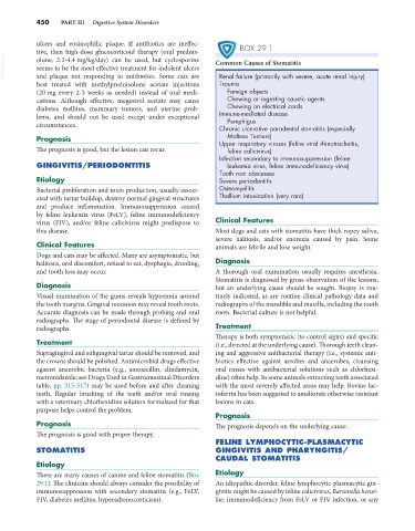
There are many causes of canine and feline stomatitis (Box Etiology
29.1). The clinician should always consider the possibility of An idiopathic disorder, feline lymphocytic-plasmacytic gin-
immunosuppression with secondary stomatitis (e.g., FeLV, givitis might be caused by feline calicivirus, Bartonella hense-
FIV, diabetes mellitus, hyperadrenocorticism). lae, immunodeficiency from FeLV or FIV infection, or any