
Page 492 - Small Animal Internal Medicine, 6th Edition
P. 492
464 PART III Digestive System Disorders
if lymphoma developed independently of the gastritis, or if
lymphoma developed from the gastritis.
VetBooks.ir cally good. Feline eosinophilic gastritis can be a component
The prognosis for canine eosinophilic gastritis is typi-
of hypereosinophilic syndrome, which typically responds
poorly to treatment. Hypereosinophilic syndrome has a
guarded to poor prognosis.
HELICOBACTER-ASSOCIATED DISEASE
Etiology
Helicobacter pylori is the principal spirochete found in
human gastric mucosa, whereas Non-H. pylori Helicobacter
(NHPH) (e.g., Helicobacter felis, Helicobacter heilmannii,
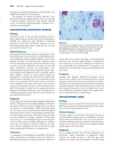
Helicobacter bizzozeronii, Helicobacter salomonis, etc.) are FIG 30.1
the principal gastric spirochetes in dogs and cats. H. pylori Air-dried smear of gastric mucosa obtained endoscopically
and stained with Diff-Quik. Numerous spirochetes are seen.
has rarely been found in cats.
The affected dog was vomiting because of an ulcerated
Clinical Features leiomyoma, and the spirochetes did not appear to be
causing disease in this animal (×1000).
Most people infected with H. pylori are asymptomatic. Those
with symptomatic H. pylori infections usually develop ulcer-
ation and gastritis with neutrophilic infiltrates. They can also people, there is no evidence that dogs or cats benefit from
develop low-grade mucosal-associated lymphoid tissue concurrent use of proton pump inhibitors. Azithromycin
(MALT) lymphoma that can be cured with antibiotic therapy and clarithromycin have been substituted for bismuth in
or gastric carcinoma. Similarly, most dogs and cats with cats. Anecdotally, some animals seem to respond to just
gastric Helicobacter infections are asymptomatic. Some erythromycin or amoxicillin. Therapy should probably last
infected animals may have nausea, hyporexia, and/or vomit- for 14 days.
ing associated with lymphocytic and occasionally neutro-
philic infiltrates. Because so many infected animals are Prognosis
asymptomatic, cause and effect have not been clearly estab- Animals with apparent Helicobacter-associated clinical
lished between Helicobacter spp. and symptomatic gastric illness seem to respond well to treatment and have a good
disease. Cats colonized with H. pylori seem to have more prognosis. However, because cause and effect are uncertain,
severe histologic lesions than those with H. felis, which in any animal not responding to therapy should be reexamined
turn may be associated with more severe lesions than those carefully, looking for other diseases. Recurrence of infection
with H. heilmannii. Currently, there is reasonable evidence after treatment commonly occurs by 6 months, but it is not
that gastric Helicobacter infections cause clinical illness (i.e., clear whether this represents a relapse of the original infec-
vomiting, hyporexia) in some dogs and cats, but there is no tion or reinfection from an outside source.
good estimate of prevalence.
PHYSALOPTERA RARA
Diagnosis
Gastric biopsy is currently required to diagnose Helicobacter Etiology
infection. The organisms are readily identified on H&E stain, Physaloptera rara is a nematode that has an indirect life cycle;
but special stains (e.g., Giemsa, Warthin-Starry) as well as beetles and crickets are the intermediate hosts. Frogs, snakes,
fluorescent in situ hybridization (FISH) make them stand mice, and birds may be paratenic hosts.
out. The bacteria are not uniformly distributed throughout
the stomach, and it is best to obtain biopsy specimens from Clinical Features
the body, fundus, and antrum. The clinician may also diag- In dogs, a single P. rara attached to the gastric mucosa can
nose this infection by cytologic evaluation of the gastric cause intractable vomiting. Cats are rarely affected, and their
mucosa (Fig. 30.1) or by looking for gastric mucosal urease clinical illness associated with P. rara is less well character-
activity. Because of the uncertain pathogenicity of Helico- ized. The vomiting usually does not resolve with antiemetics.
bacter spp., the clinician is advised to look first for other Vomitus may or may not contain bile, and affected animals
more common explanations for the animal’s clinical signs usually appear otherwise healthy.
before deciding that Helicobacter is causing disease.
Diagnosis
Treatment Ova are seldom found in feces. If fecal examinations are
A combination of metronidazole, amoxicillin, and bismuth performed, sodium dichromate or magnesium sulfate
(either subsalicylate or subcitrate) seems to be effective in solutions are usually necessary to find the eggs. Most
alleviating clinical signs in veterinary patients. Contrary to diagnoses are made when the parasites are found during