
Page 494 - Small Animal Internal Medicine, 6th Edition
P. 494
466 PART III Digestive System Disorders
VetBooks.ir
A B
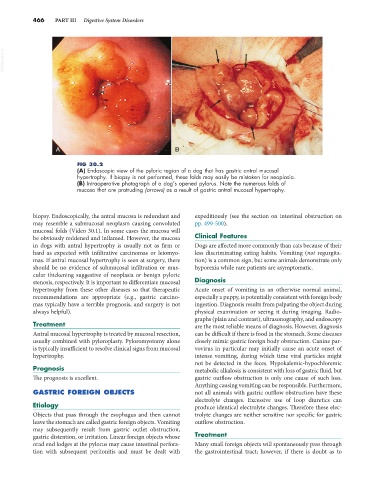
FIG 30.2
(A) Endoscopic view of the pyloric region of a dog that has gastric antral mucosal
hypertrophy. If biopsy is not performed, these folds may easily be mistaken for neoplasia.
(B) Intraoperative photograph of a dog’s opened pylorus. Note the numerous folds of
mucosa that are protruding (arrows) as a result of gastric antral mucosal hypertrophy.
biopsy. Endoscopically, the antral mucosa is redundant and expeditiously (see the section on intestinal obstruction on
may resemble a submucosal neoplasm causing convoluted pp. 499-500).
mucosal folds (Video 30.1). In some cases the mucosa will
be obviously reddened and inflamed. However, the mucosa Clinical Features
in dogs with antral hypertrophy is usually not as firm or Dogs are affected more commonly than cats because of their
hard as expected with infiltrative carcinomas or leiomyo- less discriminating eating habits. Vomiting (not regurgita-
mas. If antral mucosal hypertrophy is seen at surgery, there tion) is a common sign, but some animals demonstrate only
should be no evidence of submucosal infiltration or mus- hyporexia while rare patients are asymptomatic.
cular thickening suggestive of neoplasia or benign pyloric
stenosis, respectively. It is important to differentiate mucosal Diagnosis
hypertrophy from these other diseases so that therapeutic Acute onset of vomiting in an otherwise normal animal,
recommendations are appropriate (e.g., gastric carcino- especially a puppy, is potentially consistent with foreign body
mas typically have a terrible prognosis, and surgery is not ingestion. Diagnosis results from palpating the object during
always helpful). physical examination or seeing it during imaging. Radio-
graphs (plain and contrast), ultrasonography, and endoscopy
Treatment are the most reliable means of diagnosis. However, diagnosis
Antral mucosal hypertrophy is treated by mucosal resection, can be difficult if there is food in the stomach. Some diseases
usually combined with pyloroplasty. Pyloromyotomy alone closely mimic gastric foreign body obstruction. Canine par-
is typically insufficient to resolve clinical signs from mucosal vovirus in particular may initially cause an acute onset of
hypertrophy. intense vomiting, during which time viral particles might
not be detected in the feces. Hypokalemic-hypochloremic
Prognosis metabolic alkalosis is consistent with loss of gastric fluid, but
The prognosis is excellent. gastric outflow obstruction is only one cause of such loss.
Anything causing vomiting can be responsible. Furthermore,
GASTRIC FOREIGN OBJECTS not all animals with gastric outflow obstruction have these
electrolyte changes. Excessive use of loop diuretics can
Etiology produce identical electrolyte changes. Therefore these elec-
Objects that pass through the esophagus and then cannot trolyte changes are neither sensitive nor specific for gastric
leave the stomach are called gastric foreign objects. Vomiting outflow obstruction.
may subsequently result from gastric outlet obstruction,
gastric distention, or irritation. Linear foreign objects whose Treatment
orad end lodges at the pylorus may cause intestinal perfora- Many small foreign objects will spontaneously pass through
tion with subsequent peritonitis and must be dealt with the gastrointestinal tract; however, if there is doubt as to