
Page 499 - Small Animal Internal Medicine, 6th Edition
P. 499
CHAPTER 30 Disorders of the Stomach 471
obvious cause (e.g., NSAID or dexamethasone administra- appropriate medical therapy, resection of the ulcer(s) must
tion). Perforation may cause peritonitis and signs of an acute be considered. The stomach should be examined endoscopi-
VetBooks.ir abdomen and sepsis. Because mast cell tumors may resemble cally before surgery to determine the number and location
of the ulcers; it is surprisingly easy to miss ulcers during
almost any cutaneous lesion (especially lipomas), all cutane-
ous masses or nodules should be evaluated cytologically.
Prevention of GUE is desirable, and rational NSAID and
Hepatic failure is usually diagnosed based on the serum bio- laparotomy/gastrotomy.
chemistry profile. Contrast radiographs are diagnostic for glucocorticoid therapy are especially important. Sucral-
foreign objects but rarely demonstrate GUE. Ultrasonogra- fate (Carafate; see Table 28.5) and histamine-2 (H 2 ) recep-
phy sometimes detects gastric thickening due to infiltrated tor antagonists (see Table 28.4) have been administered to
lesions and/or mucosal defects (Fig. 30.5). Endoscopy (Video prevent GUE in dogs receiving NSAIDs, but there is no
30.3) is the most sensitive and specific tool for diagnosing good evidence that these drugs are effective prophylactic
GUE (see Figs. 27.14 to 27.17) and, in conjunction with agents. Proton pump inhibitors are effective in prevent-
biopsy, can be used to diagnose infiltrates (neoplastic or ing “stress”-induced ulceration as well as NSAID-induced
inflammatory) (see Fig. 27.16), foreign bodies (see Fig. GUE. Misoprostol (see Table 28.5) was designed to prevent
27.20), and inflammation causing GUE. Endoscopic findings NSAID-induced ulceration, and it can be used to treat ulcers.
may suggest gastrinoma if duodenal erosions are found. However, proton pump inhibitor therapy seems as effective
Serum gastrin concentrations should be measured if a gas- and has fewer side effects. No drug has been shown to be
trinoma is suspected or if there are no other likely causes. effective in preventing steroid-induced GUE (especially
dexamethasone).
Treatment
Therapy depends on the severity of GUE and whether an Prognosis
underlying cause is detected. Animals with suspected GUE The prognosis is favorable if the underlying cause can be
that is not obviously life-threatening (i.e., no evidence of controlled and if therapy prevents perforation of the ulcer.
severe anemia, shock, sepsis, severe abdominal pain, or
severe depression) may sometimes first be treated symptom-
atically if the clinician strongly suspects the cause is drug INFILTRATIVE GASTRIC DISEASES
induced or “stress” induced.
Eliminating the underlying etiology (e.g., NSAIDs, shock) NEOPLASMS
often results in resolution of the ulcer within 3 to 5 days. If Etiology
the cause is unknown or cannot be removed or if it is impor-
tant to resolve the ulcer quickly, then specific antiulcer medi- Neoplastic infiltrations (e.g., adenocarcinoma, lymphoma,
cation is appropriate. Proton pump inhibitors or sucralfate leiomyomas, leiomyosarcomas, and stromal tumors in dogs;
are the major pharmacologic options. If appropriate medical lymphoma in cats) may produce GUE through direct
therapy has not afforded clinical improvement after 5 or 6 mucosal disruption. Gastric lymphoma is typically a diffuse
days, or if the animal has life-threatening bleeding despite lesion but can produce masses. The cause and significance of
benign gastric polyps are unknown. They seem to occur
more commonly in the body and antrum.
Clinical Features
Dogs and cats with gastric tumors are usually asymptomatic
until the disease is advanced. Hyporexia (not vomiting) is the
most common initial sign. Vomiting caused by gastric neo-
plasia usually signifies advanced disease or gastric outflow
obstruction. Adenocarcinomas are typically infiltrative and
decrease emptying by impairing motility and/or obstruct-
ing the outflow tract. Weight loss is commonly caused by
nutrient loss or cancer cachexia syndrome. Hematemesis
occasionally occurs; leiomyomas seem to have the greatest
potential to cause severe, acute, upper GI bleeding. Other
bleeding gastric tumors are more likely to cause chronic
iron deficiency anemia even if GI blood loss is not obvious.
Polyps rarely cause signs unless they obstruct the pylorus.
Diagnosis
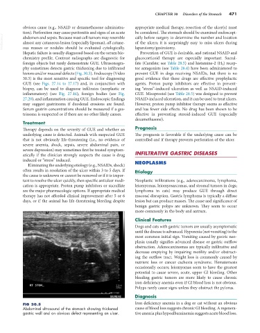
FIG 30.5 Iron deficiency anemia in a dog or cat without an obvious
Abdominal ultrasound of the stomach showing thickened cause of blood loss suggests chronic GI bleeding. A regenera-
gastric wall and an obvious defect representing an ulcer. tive anemia plus hypoalbuminemia suggests acute blood loss.