
Page 496 - Small Animal Internal Medicine, 6th Edition
P. 496
468 PART III Digestive System Disorders
VetBooks.ir
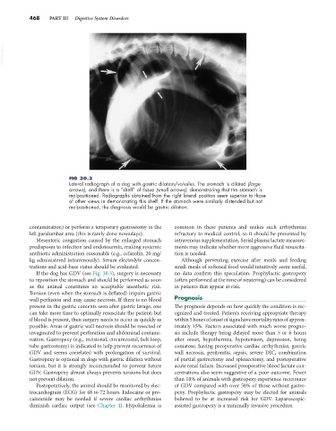
FIG 30.3
Lateral radiograph of a dog with gastric dilation/volvulus. The stomach is dilated (large
arrows), and there is a “shelf” of tissue (small arrows), demonstrating that the stomach is
malpositioned. Radiographs obtained from the right lateral position seem superior to those
of other views in demonstrating this shelf. If the stomach were similarly distended but not
malpositioned, the diagnosis would be gastric dilation.
contamination) or perform a temporary gastrostomy in the common in these patients and makes such arrhythmias
left paralumbar area (this is rarely done nowadays). refractory to medical control, so it should be prevented by
Mesenteric congestion caused by the enlarged stomach intravenous supplementation. Serial plasma lactate measure-
predisposes to infection and endotoxemia, making systemic ments may indicate whether more aggressive fluid resuscita-
antibiotic administration reasonable (e.g., cefazolin, 20 mg/ tion is needed.
kg administered intravenously). Serum electrolyte concen- Although preventing exercise after meals and feeding
trations and acid-base status should be evaluated. small meals of softened food would intuitively seem useful,
If the dog has GDV (see Fig. 30.3), surgery is necessary no data confirm this speculation. Prophylactic gastropexy
to reposition the stomach and should be performed as soon (often performed at the time of neutering) can be considered
as the animal constitutes an acceptable anesthetic risk. in patients that appear at risk.
Torsion (even when the stomach is deflated) impairs gastric
wall perfusion and may cause necrosis. If there is no blood Prognosis
present in the gastric contents seen after gastric lavage, one The prognosis depends on how quickly the condition is rec-
can take more time to optimally resuscitate the patient; but ognized and treated. Patients receiving appropriate therapy
if blood is present, then surgery needs to occur as quickly as within 5 hours of onset of signs have mortality rates of approx-
possible. Areas of gastric wall necrosis should be resected or imately 15%. Factors associated with much worse progno-
invaginated to prevent perforation and abdominal contami- sis include therapy being delayed more than 5 or 6 hours
nation. Gastropexy (e.g., incisional, circumcostal, belt loop, after onset, hypothermia, hypotension, depression, being
tube gastrostomy) is indicated to help prevent recurrence of comatose, having preoperative cardiac arrhythmias, gastric
GDV and seems correlated with prolongation of survival. wall necrosis, peritonitis, sepsis, severe DIC, combination
Gastropexy is optional in dogs with gastric dilation without of partial gastrectomy and splenectomy, and postoperative
torsion, but it is strongly recommended to prevent future acute renal failure. Increased preoperative blood lactate con-
GDV. Gastropexy almost always prevents torsions but does centrations also seem suggestive of a poor outcome. Fewer
not prevent dilation. than 10% of animals with gastropexy experience recurrence
Postoperatively, the animal should be monitored by elec- of GDV compared with over 50% of those without gastro-
trocardiogram (ECG) for 48 to 72 hours. Lidocaine or pro- pexy. Prophylactic gastropexy may be elected for animals
cainamide may be needed if severe cardiac arrhythmias believed to be at increased risk for GDV. Laparoscopic-
diminish cardiac output (see Chapter 4). Hypokalemia is assisted gastropexy is a minimally invasive procedure.