Page 524 - Small Animal Internal Medicine, 6th Edition
P. 524
496    PART III   Digestive System Disorders


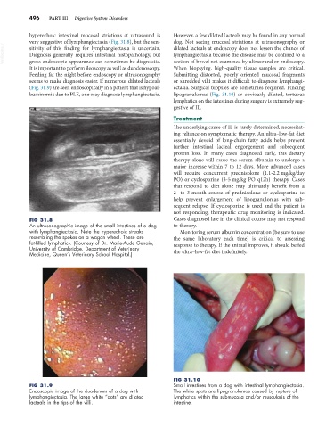
            hyperechoic intestinal mucosal striations at ultrasound is   However, a few dilated lacteals may be found in any normal
            very suggestive of lymphangiectasia (Fig. 31.8), but the sen-  dog. Not seeing mucosal striations at ultrasonography or
  VetBooks.ir  sitivity of this finding for lymphangiectasia is uncertain.   dilated lacteals at endoscopy does not lessen the chance of
                                                                 lymphangiectasia because the disease may be confined to a
            Diagnosis generally requires intestinal histopathology, but
            gross endoscopic appearance can sometimes be diagnostic.
                                                                 When  biopsying, high-quality tissue  samples  are  critical.
            It is important to perform ileoscopy as well as duodenoscopy.   section of bowel not examined by ultrasound or endoscopy.
            Feeding fat the night before endoscopy or ultrasonography   Submitting  distorted,  poorly  oriented  mucosal  fragments
            seems to make diagnosis easier. If numerous dilated lacteals   or shredded villi makes it difficult to diagnose lymphangi-
            (Fig. 31.9) are seen endoscopically in a patient that is hypoal-  ectasia. Surgical biopsies are sometimes required. Finding
            buminemic due to PLE, one may diagnose lymphangiectasia.   lipogranulomas  (Fig.  31.10) or  obviously  dilated,  tortuous
                                                                 lymphatics on the intestines during surgery is extremely sug-
                                                                 gestive of IL.
                                                                 Treatment
                                                                 The underlying cause of IL is rarely determined, necessitat-
                                                                 ing reliance on symptomatic therapy. An ultra–low-fat diet
                                                                 essentially devoid of long-chain fatty acids helps prevent
                                                                 further intestinal lacteal engorgement and subsequent
                                                                 protein  loss.  In  many cases  diagnosed  early,  this  dietary
                                                                 therapy alone will cause the serum albumin to undergo a
                                                                 major increase within 7 to 12 days. More advanced cases
                                                                 will require concurrent prednisolone (1.1-2.2 mg/kg/day
                                                                 PO) or cyclosporine (3-5 mg/kg PO q12h) therapy. Cases
                                                                 that respond to diet alone may ultimately benefit from a
                                                                 2- to 3-month course of prednisolone or cyclosporine to
                                                                 help prevent enlargement of lipogranulomas with sub-
                                                                 sequent relapse. If cyclosporine is used and the patient is
                                                                 not responding, therapeutic drug monitoring is indicated.
            FIG 31.8                                             Cases diagnosed late in the clinical course may not respond
            An ultrasonographic image of the small intestines of a dog   to therapy.
            with lymphangiectasia. Note the hyperechoic streaks    Monitoring serum albumin concentration (be sure to use
            resembling the spokes on a wagon wheel. These are    the same laboratory each time) is critical to assessing
            fat-filled lymphatics. (Courtesy of Dr. Marie-Aude Genain,   response to therapy. If the animal improves, it should be fed
            University of Cambridge, Department of Veterinary
            Medicine, Queen’s Veterinary School Hospital.)       the ultra–low-fat diet indefinitely.


                                                                 FIG 31.10
            FIG 31.9                                             Small intestines from a dog with intestinal lymphangiectasia.
            Endoscopic image of the duodenum of a dog with       The white spots are lipogranulomas caused by rupture of
            lymphangiectasia. The large white “dots” are dilated   lymphatics within the submucosa and/or muscularis of the
            lacteals in the tips of the villi.                   intestine.

