
Page 529 - Small Animal Internal Medicine, 6th Edition
P. 529
CHAPTER 31 Disorders of the Intestinal Tract 501
linear foreign object has been present a long time, it may nography is quick and reasonably sensitive and specific for
embed itself in the intestinal mucosa, making intestinal detecting intussusceptions (see Fig. 27.8, B). Colonoscopy
VetBooks.ir resection necessary. When massive intestinal resection is can be definitive if the intussuscepted intestine is seen ex-
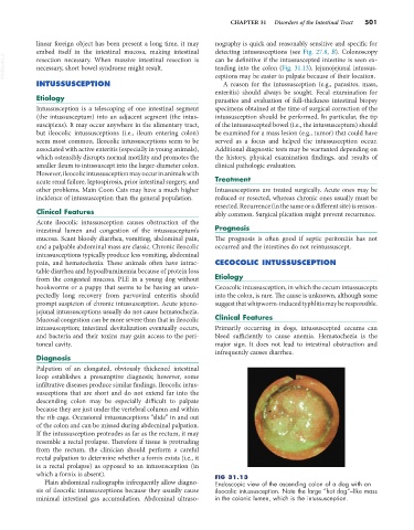
tending into the colon (Fig. 31.13). Jejunojejunal intussus-
necessary, short bowel syndrome might result.
INTUSSUSCEPTION ceptions may be easier to palpate because of their location.
A reason for the intussusception (e.g., parasites, mass,
enteritis) should always be sought. Fecal examination for
Etiology parasites and evaluation of full-thickness intestinal biopsy
Intussusception is a telescoping of one intestinal segment specimens obtained at the time of surgical correction of the
(the intussusceptum) into an adjacent segment (the intus- intussusception should be performed. In particular, the tip
suscipiens). It may occur anywhere in the alimentary tract, of the intussuscepted bowel (i.e., the intussusceptum) should
but ileocolic intussusceptions (i.e., ileum entering colon) be examined for a mass lesion (e.g., tumor) that could have
seem most common. Ileocolic intussusceptions seem to be served as a focus and helped the intussusception occur.
associated with active enteritis (especially in young animals), Additional diagnostic tests may be warranted depending on
which ostensibly disrupts normal motility and promotes the the history, physical examination findings, and results of
smaller ileum to intussuscept into the larger-diameter colon. clinical pathologic evaluation.
However, ileocolic intussusception may occur in animals with
acute renal failure, leptospirosis, prior intestinal surgery, and Treatment
other problems. Main Coon Cats may have a much higher Intussusceptions are treated surgically. Acute ones may be
incidence of intussusception than the general population. reduced or resected, whereas chronic ones usually must be
resected. Recurrence (in the same or a different site) is reason-
Clinical Features ably common. Surgical plication might prevent recurrence.
Acute ileocolic intussusception causes obstruction of the
intestinal lumen and congestion of the intussusceptum’s Prognosis
mucosa. Scant bloody diarrhea, vomiting, abdominal pain, The prognosis is often good if septic peritonitis has not
and a palpable abdominal mass are classic. Chronic ileocolic occurred and the intestines do not reintussuscept.
intussusceptions typically produce less vomiting, abdominal
pain, and hematochezia. These animals often have intrac- CECOCOLIC INTUSSUSCEPTION
table diarrhea and hypoalbuminemia because of protein loss
from the congested mucosa. PLE in a young dog without Etiology
hookworms or a puppy that seems to be having an unex- Cecocolic intussusception, in which the cecum intussuscepts
pectedly long recovery from parvoviral enteritis should into the colon, is rare. The cause is unknown, although some
prompt suspicion of chronic intussusception. Acute jejuno- suggest that whipworm-induced typhlitis may be responsible.
jejunal intussusceptions usually do not cause hematochezia.
Mucosal congestion can be more severe than that in ileocolic Clinical Features
intussusception; intestinal devitalization eventually occurs, Primarily occurring in dogs, intussuscepted cecums can
and bacteria and their toxins may gain access to the peri- bleed sufficiently to cause anemia. Hematochezia is the
toneal cavity. major sign. It does not lead to intestinal obstruction and
infrequently causes diarrhea.
Diagnosis
Palpation of an elongated, obviously thickened intestinal
loop establishes a presumptive diagnosis; however, some
infiltrative diseases produce similar findings. Ileocolic intus-
susceptions that are short and do not extend far into the
descending colon may be especially difficult to palpate
because they are just under the vertebral column and within
the rib cage. Occasional intussusceptions “slide” in and out
of the colon and can be missed during abdominal palpation.
If the intussusception protrudes as far as the rectum, it may
resemble a rectal prolapse. Therefore if tissue is protruding
from the rectum, the clinician should perform a careful
rectal palpation to determine whether a fornix exists (i.e., it
is a rectal prolapse) as opposed to an intussusception (in
which a fornix is absent). FIG 31.13
Plain abdominal radiographs infrequently allow diagno- Endoscopic view of the ascending colon of a dog with an
sis of ileocolic intussusceptions because they usually cause ileocolic intussusception. Note the large “hot dog”–like mass
minimal intestinal gas accumulation. Abdominal ultraso- in the colonic lumen, which is the intussusception.