
Page 528 - Small Animal Internal Medicine, 6th Edition
P. 528
500 PART III Digestive System Disorders
object usually prevent the intestines from dilating. Plain if it will now pass through the intestines without further
radiographs may reveal small gas bubbles in the intestines, problem. Surgery is indicated if the animal does not appear
VetBooks.ir especially in the region of the duodenum, and obvious better 12 to 24 hours after the object is cut free from its point
of fixation.
intestinal pleating may occasionally be seen (Fig. 31.12). If
If there is doubt as to the length of time the object has
contrast radiographs are performed (use an isotonic iodine
contrast agent), they typically reveal a pleated or bunched been present or if it is fixed at the pylorus, surgery is usually
intestinal pattern, which is diagnostic of linear foreign body. a safer therapeutic approach. Endoscopic removal occasion-
These objects are sometimes seen endoscopically, lodged at ally succeeds, but the clinician must be careful because it is
the pylorus. easy to rupture devitalized intestine and cause peritonitis. If
the clinician can pass the tip of the endoscope to near the
Treatment aborad end of the object and pull it out by grabbing the
Abdominal surgery is typically required to remove linear aborad end, surgery is sometimes unnecessary.
foreign objects. If the animal is otherwise healthy, if the
linear foreign object has been present for only 1 or 2 days, Prognosis
and if it is fixed under the tongue, the object may be cut The prognosis is usually good if severe septic peritonitis is
loose from its attachment at the base of the tongue to see absent and massive intestinal resection is unnecessary. If a
A
C
B
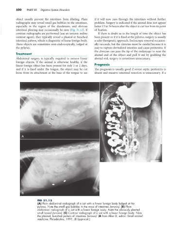
FIG 31.12
(A) Plain abdominal radiograph of a cat with a linear foreign body lodged at the
pylorus. Note the small gas bubbles in the mass of intestines (arrows). (B) Plain
abdominal radiograph of a cat with a linear foreign body. Note the obviously pleated
small bowel (arrows). (C) Contrast radiograph of a cat with a linear foreign body. Note
the pleated, bunched pattern of intestines (arrows). (A from Allen D, editor: Small animal
medicine, Philadelphia, 1991, JB Lippincott.)