
Page 624 - Small Animal Internal Medicine, 6th Edition
P. 624
596 PART IV Hepatobiliary and Exocrine Pancreatic Disorders
hepatitis. These dogs usually present at about 4 years of age, Diagnosis
but may be younger (Fig. 36.6). Eventually, if not treated, The magnitude of increase in liver enzyme activities and the
VetBooks.ir affected dogs will develop cirrhosis. storage disease are very similar to those of dogs with idio-
diagnostic imaging findings in dogs with chronic copper
The clinical signs and progression in other breeds with
pathic chronic hepatitis. Therefore a definitive diagnosis
copper storage disease are similar to those in Bedlington
Terriers. The disease in Dalmatians is associated with acute requires a liver biopsy and determination or estimation
onset, rapid progression, and very high levels of hepatic of the copper concentration in the liver. This can be done
copper in the absence of significant clinical, clinicopatho- qualitatively on formalin-fixed sections using rhodanine or
logic, or histologic evidence of cholestasis. Affected dogs rubeanic acid staining to detect copper; correlations between
usually present as young adults with acute onset of GI signs the quantitative and qualitative estimations of copper accu-
and PU-PD, by which time severe liver disease is already mulation have been published (Shih et al., 2007). Digital
present. Labrador Retrievers with copper storage disease scanning of rhodanine-stained sections is a more objective
have an average age at presentation of 7 to 9 years (range, qualitative measure of hepatic copper (Center et al., 2013).
2.5–14 years). The clinical signs are relatively mild and The finding of large accumulations of copper in hepatocytes
include anorexia, vomiting, and lethargy. Doberman Pin- on cytology with rubeanic acid is also suggestive of copper
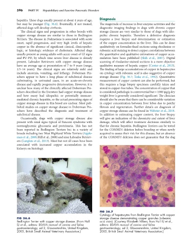
schers appear to have a long phase of subclinical disease storage disease (Fig. 36.7; Teske et al., 1992). Quantitative
culminating, in untreated cases, in an acute-on-chronic measurement of copper content can also be performed, but
disease and rapidly progressive deterioration. However, it is this requires a large biopsy specimen carefully taken and
unclear how many of the clinically affected Doberman Pin- stored in copper-free tubes. The concentration of copper that
schers described in the literature had copper storage disease is considered pathologic is controversial but > 1000 µg/g dry
and how many had idiopathic or potentially immune- weight liver is generally considered significant. The clinician
mediated chronic hepatitis, so the actual presenting signs of should also be aware that there can be considerable variation
copper storage disease in this breed are unclear. Most pub- in copper concentration between liver lobes due to patchy
lished studies on copper storage disease in Doberman Pin- fibrosis and regeneration. Further details on diagnosis of
schers have described the diagnosis and treatment of copper storage disease can be found in Webster et al., 2019.
subclinical disease. In addition to estimating copper content, the liver biopsy
Occasionally, dogs with copper storage disease also will give an indication of the chronicity and extent of liver
present with renal signs typical of Fanconi syndrome with damage, which will affect treatment decisions similarly to
normoglycemic glycosuria and proteinuria. This has not that for chronic hepatitis. Bedlington Terriers can be tested
been reported in Bedlington Terriers but in a variety of for the COMMD1 deletion before breeding or when newly
breeds including two West Highland White Terriers (Apple- acquired to assess their risk for this disease, but an absence
man et al., 2008; Hill et al., 2008) and nine Labrador Retriev- of the COMMD1 deletion does not guarantee that the dog
ers (Langlois et al., 2013). Most but not all cases have been
associated with increased copper accumulation in the
kidneys on histology.
FIG 36.7
Cytology of hepatocytes from Bedlington Terrier with copper
FIG 36.6 storage disease demonstrating copper granules (rubeanic
Bedlington Terrier with copper storage disease. (From Hall acid stain). (Courtesy Elizabeth Villiers; from Hall EJ et al.,
EJ et al., editors: BSAVA manual of canine and feline editors: BSAVA manual of canine and feline
gastroenterology, ed 2, Gloucestershire, United Kingdom, gastroenterology, ed 2, Gloucestershire, United Kingdom,
2005, British Small Animal Veterinary Association.) 2005, British Small Animal Veterinary Association.)