
Page 628 - Small Animal Internal Medicine, 6th Edition
P. 628
600 PART IV Hepatobiliary and Exocrine Pancreatic Disorders
similar to those in cats with neutrophilic cholangitis (see gallbladder wall visible radiographically or ultrasonographi-
Chapter 35). Dogs can be of any age or breed, and the typical cally. Antibiotic resistance is relatively common among
VetBooks.ir presentation is acute onset of anorexia, jaundice, and vomit- isolates and can also develop during therapy, underscoring
the importance of obtaining bile samples for culture and
ing, with or without pyrexia, although not all affected dogs
are jaundiced. In some cases there may have been a previous
association with cholecystitis or cholangitis; the cause and
history of acute enteritis or pancreatitis, suggesting a poten- sensitivity whenever possible. Choleliths can be found in
tial cause for ascending biliary infection from the gut. effect relationship is not always clear.
Mechanical obstruction and gallbladder mucocele (see later) Treatment involves 4 to 6 weeks of antibiotics, preferably
should be ruled out first, usually by ultrasonography, and based on the results of culture and sensitivity, together with
then liver and bile and/or gallbladder mucosa specimens antioxidants and choleretics. As in cats, amoxicillin is a good
should be obtained for histopathology and microbial culture initial choice at a dose of 15 to 20 mg/kg PO q8h combined
and sensitivity testing, preferably before antibiotic treatment with ursodeoxycholic acid at a dose of 15 mg/kg PO every
is initiated. Concurrent cholecystitis is common in those 24 hours or split into two doses q12 hours. One recent study
cases in which it is investigated (Harrison et al., 2018). showed cholecystectomy reduced the risk of death in affected
Liver biopsies and bile samples can be obtained by direct dogs, so this should be seriously considered, particularly
visualization during surgery, laparoscopy, or ultrasono- when gallbladder abnormalities are found on ultrasound
graphic guidance. The latter method carries a greater risk of (Harrison et al., 2018).
bile leakage; to minimize this, a 22-gauge needle attached to a
12-mL syringe is used for cholecystocentesis (bile retrieval), GALLBLADDER MUCOCELE
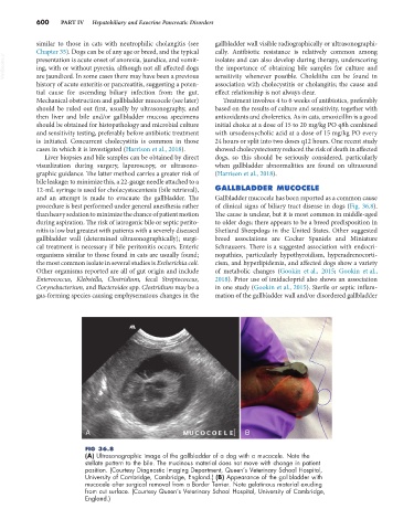
and an attempt is made to evacuate the gallbladder. The Gallbladder mucocele has been reported as a common cause
procedure is best performed under general anesthesia rather of clinical signs of biliary tract disease in dogs (Fig. 36.8).
than heavy sedation to minimize the chance of patient motion The cause is unclear, but it is most common in middle-aged
during aspiration. The risk of iatrogenic bile or septic perito- to older dogs; there appears to be a breed predisposition in
nitis is low but greatest with patients with a severely diseased Shetland Sheepdogs in the United States. Other suggested
gallbladder wall (determined ultrasonographically); surgi- breed associations are Cocker Spaniels and Miniature
cal treatment is necessary if bile peritonitis occurs. Enteric Schnauzers. There is a suggested association with endocri-
organisms similar to those found in cats are usually found; nopathies, particularly hypothyroidism, hyperadrenocorti-
the most common isolate in several studies is Escherichia coli. cism, and hyperlipidemia, and affected dogs show a variety
Other organisms reported are all of gut origin and include of metabolic changes (Gookin et al., 2015; Gookin et al.,
Enterococcus, Klebsiella, Clostridium, fecal Streptococcus, 2018). Prior use of imidacloprid also shows an association
Corynebacterium, and Bacteroides spp. Clostridium may be a in one study (Gookin et al., 2015). Sterile or septic inflam-
gas-forming species causing emphysematous changes in the mation of the gallbladder wall and/or disordered gallbladder
A B
FIG 36.8
(A) Ultrasonographic image of the gallbladder of a dog with a mucocele. Note the
stellate pattern to the bile. The mucinous material does not move with change in patient
position. (Courtesy Diagnostic Imaging Department, Queen’s Veterinary School Hospital,
University of Cambridge, Cambridge, England.) (B) Appearance of the gallbladder with
mucocele after surgical removal from a Border Terrier. Note gelatinous material exuding
from cut surface. (Courtesy Queen’s Veterinary School Hospital, University of Cambridge,
England.)